
Congenital hereditary endothelial dystrophy (CHED) is a rare inherited corneal endothelial disorder.
It presents at or shortly after birth with bilateral, diffuse corneal clouding. It is one of a small handful of conditions that must be urgently distinguished from congenital glaucoma. This applies to any infant with cloudy corneas.
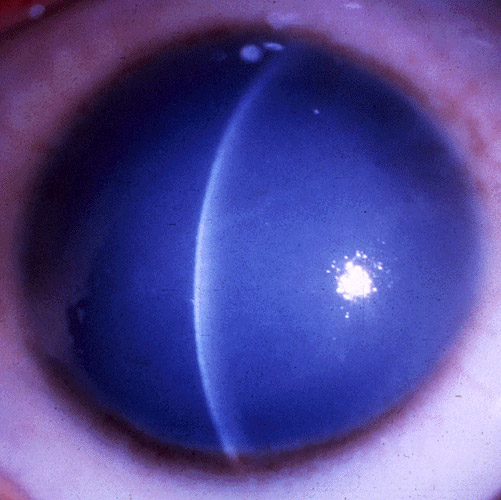
This distinction carries enormous practical stakes. The two conditions require entirely different management — genuinely time-sensitive in the case of glaucoma.
Genetics
CHED is now understood to represent a spectrum related to mutations in SLC4A11.
Autosomal recessive inheritance is the form most commonly encountered, and generally the more severe. It presents at birth with dense, opaque corneal clouding.
Older classification schemes described a milder, autosomal dominant form with later onset and slower progression.
However, modern genetic understanding has clarified something important. The great majority of clinically significant cases seen in practice are the recessive, congenital-onset form.
Pathophysiology
The underlying defect impairs normal corneal endothelial pump function from birth.
This prevents the endothelium from adequately clearing fluid from the stroma, as a normal endothelial layer would. The result is diffuse, bilateral stromal and epithelial edema present from the earliest days of life.
This gives the cornea a hazy, ground-glass appearance, rather than its normal clarity. Fuchs dystrophy, discussed in its own dedicated article, develops from progressive endothelial cell loss over decades.
CHED is different: it reflects a fundamental developmental failure present from the start.
All-fit Slit-Lamp Adapter
Record and share exactly what you see at the slit lamp. One adapter fits any slit lamp or surgical microscope — and any smartphone.
From Choroida — the team behind this siteClinical Presentation
- Bilateral, diffuse corneal clouding present at birth or developing within the first weeks of life, often symmetric between the two eyes
- Corneal thickness is significantly increased (unlike the normal or near-normal corneal thickness typically seen in congenital glaucoma, where the primary structural change is corneal enlargement and Descemet membrane tears rather than diffuse edema)
- Photophobia and tearing, though generally less pronounced than the marked photophobia and blepharospasm classically associated with congenital glaucoma
- Nystagmus, often developing secondary to the poor, blurred visual input the child receives from the clouded corneas during the critical period of visual development
- Notably normal intraocular pressure and normal corneal diameter — this is the single most important distinguishing feature from congenital glaucoma, and confirming it is the central task of the initial evaluation
Distinguishing CHED From Congenital Glaucoma
Congenital glaucoma produces corneal clouding through a fundamentally different mechanism.
Elevated intraocular pressure causes corneal enlargement (buphthalmos) and Descemet’s membrane breaks (Haab’s striae), letting aqueous edematize the stroma.
It is accompanied by an enlarged corneal diameter and elevated intraocular pressure on measurement. Photophobia, tearing, and blepharospasm are also often more prominent than typically seen in CHED.
Congenital glaucoma is a genuine emergency, requiring prompt surgical intervention to prevent irreversible optic nerve damage.
Any infant presenting with cloudy corneas needs intraocular pressure measurement and careful corneal diameter assessment as an urgent priority. This often requires examination under anesthesia, given how difficult a reliable, cooperative exam is in an infant.
This should happen before CHED can be comfortably assumed as the explanation.
Differential Diagnosis of Neonatal Corneal Clouding
- Congenital glaucoma — elevated intraocular pressure, corneal enlargement, Haab’s striae, discussed in its own dedicated article on this site; the critical alternative diagnosis to exclude urgently
- Birth trauma (forceps injury) — typically unilateral or asymmetric, with a clear history of a difficult or instrumented delivery, and often vertical or oblique Descemet membrane tears on careful exam
- Sclerocornea — a congenital anomaly in which the cornea takes on an opaque, sclera-like quality, generally more peripheral and with a different overall appearance than the diffuse, central-predominant clouding of CHED, discussed in its own dedicated article on this site
- Metabolic and storage disorders (mucopolysaccharidoses and others) — generally accompanied by other systemic findings that help distinguish them from an isolated corneal dystrophy, and worth considering when corneal clouding is accompanied by additional developmental or systemic abnormalities
- Congenital corneal infection (herpes simplex, rubella) — a relevant maternal and perinatal history usually points toward this category rather than an inherited dystrophy.
Management
Because CHED significantly impairs visual input from birth, there is a real and urgent risk of severe amblyopia.
This applies if the corneal clouding is not addressed during the critical period of early visual development.
Prompt intervention, rather than a “wait and see” approach, is generally appropriate.
This holds once the diagnosis is established and congenital glaucoma has been confidently excluded. Corneal transplantation is the definitive treatment.
This is traditionally penetrating keratoplasty, though endothelial keratoplasty techniques are increasingly explored in selected pediatric cases.
It is performed as early as reasonably feasible, given the amblyogenic stakes. However, pediatric corneal transplantation carries its own well-recognized surgical and long-term challenges.
These include a higher rejection risk than adult keratoplasty, and the need for aggressive, coordinated amblyopia management after surgery.
Genetic counseling for the family addresses recurrence risk given the syndrome’s typically autosomal recessive inheritance. Long-term, coordinated pediatric ophthalmic follow-up is essential for optimizing the child’s ultimate visual outcome.
This addresses not just the corneal graft itself, but the amblyopia, refractive error, and any associated nystagmus.


Document what you see
Two smartphone imaging tools built for everyday clinic use — one for the slit lamp, one for the fundus.
From Choroida — the team behind this siteReferences
- Kumar A, Bhattacharjee S, Prakash DR, Sadanand CS. Genetic analysis of two Indian families affected with congenital hereditary endothelial dystrophy: two novel mutations in SLC4A11. Molecular Vision.
- Weiss JS, Møller HU, Aldave AJ, et al. IC3D classification of corneal dystrophies — edition 2. Cornea.
- American Academy of Ophthalmology. Basic and Clinical Science Course, Section 6: Pediatric Ophthalmology and Strabismus.
