
Case Study
A 58-year-old hyperopic man presented with gradual, painless blurring of vision in his right eye over six months. He denied flashes, floaters, or field defects.
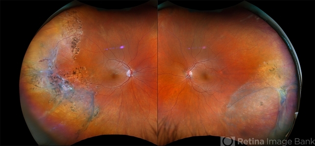
Visual acuity was 20/30 in the right eye and 20/20 in the left. Fundus examination revealed a smooth, dome-shaped, translucent elevation of the inferotemporal retina extending toward the macula, with an underlying clear cavity.
The surface vessels appeared immobile and elevated, and no pigment demarcation line or retinal tears were observed.
Optical coherence tomography (OCT) demonstrated a splitting of the neurosensory retina at the outer plexiform layer with a schitic cavity, confirming bullous retinoschisis.
The patient was managed conservatively with regular follow-up, as there was no sign of progression or associated retinal detachment.
Disease Entity
Bullous Retinoschisis is an acquired degenerative condition characterized by a splitting of the retinal layers, typically occurring within the outer plexiform or inner nuclear layers.
The term “bullous” refers to its smooth, dome-shaped elevation that mimics a localized, non-rhegmatogenous retinal detachment.
It most commonly affects the inferotemporal quadrant of the peripheral retina and is often asymptomatic, discovered incidentally during routine examination.
However, in some cases, it may extend posteriorly or be complicated by retinal holes, leading to vision-threatening detachments.
Fundus Explorer Pro
Photograph the retinal findings described here with the phone already in your pocket — 22 D optics and built-in illumination in one handheld unit.
From Choroida — the team behind this sitePathophysiology
The fundamental mechanism of bullous retinoschisis involves degenerative microcystic changes within the retina that coalesce and form cavities between layers.
These changes result in splitting of the retina, most often at the junction between the outer plexiform and inner nuclear layers, or less commonly at the nerve fiber layer.
The process is thought to arise from:
-
Age-related microstructural degeneration of the retina.
-
Vitreous traction or microglial activation which causes fluid accumulation within the retinal layers.
-
Genetic predisposition, as seen in X-linked juvenile retinoschisis due to mutations in the RS1 gene (inherited form).
Fluid within the schisis cavity is typically proteinaceous and acellular, distinguishing it from the subretinal fluid seen in rhegmatogenous retinal detachment (RRD).
The retinal vessels remain taut and immobile over the elevated area, a clinical hallmark that helps differentiate bullous retinoschisis from other retinal elevations.
Epidemiology
-
Age group: Most commonly affects individuals over 50 years of age.
-
Gender: Slight male predominance.
-
Refractive status: Frequently associated with hyperopia.
-
Laterality: Often bilateral, though usually asymmetric.
-
Prevalence: Reported in approximately 4–7% of adults undergoing routine fundus examination.
Clinical Features
Bullous retinoschisis is usually asymptomatic unless the schisis involves the macula or is complicated by retinal detachment.
Symptoms (if present):
-
Gradual blurring or distortion of vision.
-
Peripheral visual field defects.
-
Rarely, acute vision loss if associated with outer or inner layer breaks leading to detachment.
Fundus findings:
-
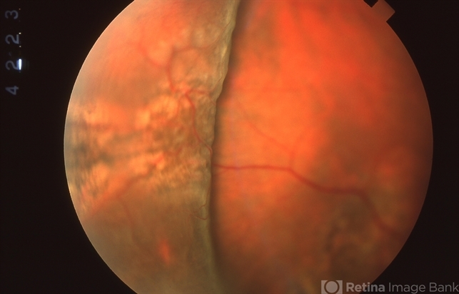
A smooth, dome-shaped, translucent retinal elevation, typically in the inferotemporal quadrant.
-
The retinal surface vessels appear immobile and follow the curvature of the elevation.
-
No undulating movement of the elevated retina on eye movement (unlike retinal detachment).
-
Occasionally, small inner or outer layer holes can be observed within the schisis cavity.
OCT findings:
-
Splitting of the retina, most often at the outer plexiform layer.
-
Smoothly elevated retina with a clear schisis cavity.
-
Absence of subretinal fluid.
-
Intact retinal pigment epithelium (RPE) and photoreceptor layer unless detachment develops.

Differential Diagnosis
Distinguishing bullous retinoschisis from rhegmatogenous retinal detachment (RRD) is critical, as management differs significantly.
| Feature | Bullous Retinoschisis | Rhegmatogenous Retinal Detachment |
|---|---|---|
| Surface appearance | Smooth, taut, immobile | Corrugated, mobile |
| Vessels | Elevated and immobile | Mobile and torturous |
| Demarcation line | Absent | May be present |
| Subretinal fluid | None | Present |
| Photocoagulation reaction | None (retina does not whiten) | Retinal whitening occurs |
| OCT finding | Retinal splitting (schisis cavity) | Separation of the neurosensory retina from the RPE |
Other differential diagnoses include:
-
X-linked juvenile retinoschisis
-
Cystoid macular edema
-
Retinal pigment epithelial detachment
-
Retinoschisis-related retinal detachment
Examination Findings
Funduscopy:
-
Dome-shaped, transparent retinal elevation without shifting fluid.
-
Taut retinal vessels bridging the lesion.
-
Absence of pigmentary changes or retinal folds.
OCT:
-
Confirms the schisis cavity between retinal layers.
-
May show both inner and outer layer holes.
Visual field testing:
-
Absolute scotoma corresponding to the schisis area (as opposed to relative scotoma in RRD).
B-scan ultrasonography:
-
Shows smooth retinal elevation with low reflectivity and no subretinal fluid.
Diagnosis
Diagnosis is primarily clinical and imaging-based, utilizing:
-
Indirect ophthalmoscopy for fundus evaluation.
-
Optical Coherence Tomography (OCT) to identify the layer of retinal splitting.
-
Fundus Autofluorescence (FAF), which may show mild hypoautofluorescence corresponding to schisis areas.
-
Fluorescein Angiography (FA), usually normal, confirming the absence of leakage.
Complications
While most cases remain stable, a minority may develop serious complications:
-
Outer layer holes: Allow fluid movement into the subretinal space.
-
Retinal detachment: Occurs if both inner and outer layers break, coexist, permitting communication between the vitreous cavity and subretinal space.
-
Macular involvement: May cause reduced central vision.
Management
Most cases of bullous retinoschisis are benign and non-progressive, requiring observation rather than intervention.
Observation:
-
Regular follow-up every 6–12 months with fundus and OCT examinations.
-
Patient education to report sudden vision changes or visual field defects.
Laser Photocoagulation:
-
It may be applied around outer-layer holes near the posterior margin to prevent retinal detachment in high-risk cases.
Surgical Intervention:
-
Pars plana vitrectomy (PPV) or scleral buckle may be considered if retinal detachment develops due to coexisting breaks.
-
Surgery aims to close both the inner and outer layer holes and reattach the retina.
Prognosis:
-
Excellent in uncomplicated cases; most remain stable for years.
-
Visual prognosis worsens if macula-involving schisis or detachment occurs.
Prevention and Follow-Up
-
Regular monitoring for retinal holes or signs of progression.
-
Avoid excessive ocular trauma.
-
Manage associated systemic conditions like hypertension or hyperopia.
-
Annual dilated fundus exams for both eyes.


Document what you see
Two smartphone imaging tools built for everyday clinic use — one for the slit lamp, one for the fundus.
From Choroida — the team behind this siteReferences
-
Byer NE. Long-term natural history study of senile retinoschisis with implications for management. Ophthalmology. 1986;93(9):1127–1137.
-
Shimada N, Ohno-Matsui K, et al. Retinoschisis and its clinical features: imaging findings and natural course. Am J Ophthalmol. 2006;141(1):128–134.
-
Futterman S, et al. Degenerative retinoschisis and its differentiation from retinal detachment. Surv Ophthalmol. 2008;53(5):471–489.
-
Lee WK, et al. Spectral-domain OCT features of bullous retinoschisis and associated complications. Br J Ophthalmol. 2014;98(10):1325–1331.
Test yourself
A few questions straight from this article.
-
At which retinal layer does the splitting of bullous retinoschisis most often occur?
Bullous retinoschisis splits the neurosensory retina most often at the outer plexiform layer, or at the junction with the inner nuclear layer; nerve fiber layer splitting is less common. -
Which retinal quadrant is most commonly affected by bullous retinoschisis?
The lesion characteristically occupies the inferotemporal peripheral retina and is often found incidentally on routine examination. -
What is the reported prevalence of bullous retinoschisis among adults having routine fundus examination?
Around 4-7% of adults undergoing routine fundus examination are found to have retinoschisis, most often after the age of 50 and frequently bilaterally. -
Which refractive status is most frequently associated with bullous retinoschisis?
Bullous retinoschisis is frequently associated with hyperopia, affects people over 50 with a slight male predominance, and is often bilateral though asymmetric. -
Which fundus sign helps separate bullous retinoschisis from rhegmatogenous retinal detachment?
In retinoschisis the elevation is smooth and taut with immobile vessels and no undulation, whereas rhegmatogenous detachment is corrugated and mobile. -
What type of visual field defect corresponds to the area of bullous retinoschisis?
Retinoschisis produces an absolute scotoma over the affected area, in contrast to the relative scotoma found in rhegmatogenous retinal detachment. -
How does the elevated retina in bullous retinoschisis respond to test photocoagulation?
The schitic retina does not whiten on photocoagulation, whereas detached retina in rhegmatogenous detachment shows retinal whitening. -
Mutation of which gene causes the inherited X-linked juvenile form of retinoschisis?
The inherited form, X-linked juvenile retinoschisis, results from mutations in the RS1 gene, whereas the bullous type is usually an acquired degenerative process. -
What is the recommended management of uncomplicated, non-progressive bullous retinoschisis?
Most cases are benign and non-progressive, so observation with fundus and OCT review every 6-12 months plus patient education about sudden visual change is sufficient. -
What combination of breaks leads to retinal detachment complicating bullous retinoschisis?
Detachment occurs when inner and outer layer breaks coexist, allowing communication between the vitreous cavity and the subretinal space; surgery aims to close both.
