
Silent Sinus Syndrome is one of the few orbital conditions a patient notices before the clinician does.
The eye simply looks sunken, and it happens so slowly that photographs are often the first evidence anything has changed.
What makes it deceptive is the name: the maxillary sinus is collapsing, yet there is no pain, no nasal blockage, and no history of sinusitis.
For clinicians, recognizing Silent Sinus Syndrome—also called chronic maxillary atelectasis or the imploding antrum syndrome—matters because it is surgically correctable, and because it is easily mistaken for trauma or for proptosis of the opposite eye.
A single CT scan settles the diagnosis, so the whole challenge is remembering to think of it.
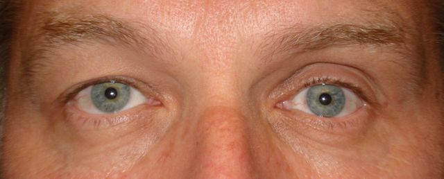
What Is Silent Sinus Syndrome?
Silent Sinus Syndrome is a painless, progressive unilateral enophthalmos and hypoglobus caused by chronic collapse of the maxillary sinus.
The defining combination is:
- Gradual, painless enophthalmos on one side
- Downward displacement of the globe (hypoglobus)
- An opacified, shrunken maxillary sinus with an inwardly bowed orbital floor
- Absence of symptoms of sinus disease
It is the silence—no pain, no discharge, no congestion—that separates it from ordinary chronic sinusitis.
Epidemiology
The condition is uncommon and almost always unilateral.
- Most patients present between the third and fifth decades
- There is no clear sex predilection
- Bilateral cases are exceptional
Because it is rare and painless, many patients are reassured for months before the diagnosis is made.
All-fit Slit-Lamp Adapter
Record and share exactly what you see at the slit lamp. One adapter fits any slit lamp or surgical microscope — and any smartphone.
From Choroida — the team behind this sitePathophysiology
The whole sequence begins with a blocked maxillary sinus outflow tract.
The chain of events is:
- Obstruction of the sinus ostium, often by a lateralized uncinate process
- Hypoventilation of the sinus and gradual resorption of trapped gas
- Persistent negative pressure within the sinus
- Inward retraction and thinning of the sinus walls, especially the orbital floor
- Expansion of the bony orbital volume
Because the globe occupies a fixed volume, an enlarged orbit inevitably means the eye sits further back and lower.
Clinical Presentation
Symptoms
The complaint is almost always cosmetic rather than visual.
- A “sunken” or “smaller” eye noticed by the patient, family, or from old photographs
- Facial asymmetry
- No pain, no nasal obstruction, and no discharge
- Vision is typically normal, and diplopia is uncommon
Examination Findings
The signs are subtle in isolation but characteristic in combination.
- Enophthalmos: usually 2–5 mm, best appreciated from above or below rather than face-on
- Hypoglobus: the affected globe sits lower than its fellow
- Deep superior sulcus from posterior displacement of orbital fat
- Upper eyelid retraction or pseudoptosis with an asymmetric palpebral fissure
- Malar flattening and midfacial asymmetry
- Ocular motility and visual acuity are usually intact
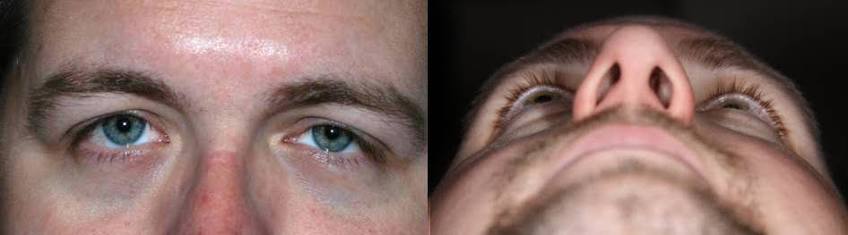
Viewing the patient from below—the worm’s-eye view—is the quickest way to confirm enophthalmos that is not obvious from the front.
Why Silent Sinus Syndrome Is So Often Missed
Almost every delay in diagnosis follows the same few patterns.
- The absence of sinus symptoms steers attention away from the sinuses entirely
- The asymmetry is misread as proptosis of the opposite eye rather than enophthalmos of the affected one
- It is wrongly attributed to remote facial trauma, since an old orbital floor fracture looks similar
- The change is so gradual that neither patient nor clinician can date its onset
Whenever one eye looks sunken without a clear history of trauma, the maxillary sinus deserves a look.
Diagnostic Evaluation
The diagnosis is clinical in suspicion and radiological in confirmation.
Clinical Assessment
- Exophthalmometry to quantify the difference between the two eyes
- Measurement of vertical globe position to document hypoglobus
- Comparison with old photographs to establish the time course
CT Imaging
Non-contrast CT of the orbits and sinuses is diagnostic and should include coronal views.
- Opacified maxillary sinus with clearly reduced volume
- Inferior bowing and thinning of the orbital floor
- Retraction of the other sinus walls toward the centre of the antrum
- Lateralized uncinate process with an obstructed infundibulum
- Increased orbital volume with an enlarged inferior orbital fat compartment
The combination of a small, opacified antrum and a downwardly bowed orbital floor is effectively pathognomonic.
Differential Diagnosis
Several conditions produce unilateral enophthalmos or the appearance of it:
- Old orbital floor fracture — the closest mimic, distinguished by trauma history and fracture morphology on CT
- Contralateral proptosis, for example thyroid-associated ophthalmopathy — the classic pseudo-enophthalmos trap
- Metastatic scirrhous carcinoma (notably breast) — causes enophthalmos through fibrosis
- Parry–Romberg syndrome and linear scleroderma — progressive hemifacial atrophy
- Orbital fat atrophy after radiotherapy or surgery
- Congenital facial asymmetry
CT resolves nearly all of these, which is why imaging should not be delayed once enophthalmos is confirmed.
Management
Treatment is surgical and best delivered jointly by otolaryngology and oculoplastics.
Restoring Sinus Ventilation
- Functional endoscopic sinus surgery with uncinectomy and middle meatal antrostomy
- This relieves the negative pressure and halts further collapse
- It is the essential first step in every case
Orbital Floor Reconstruction
- Indicated for residual enophthalmos or hypoglobus that remains cosmetically significant
- Performed with an orbital floor implant to restore globe position
- Not every patient requires it
Timing
- A staged approach is commonly preferred, with orbital reconstruction deferred for several months
- Delay allows any spontaneous improvement after re-ventilation to declare itself
- Simultaneous surgery is an option in selected cases
Restoring ventilation stops the process; restoring the orbital floor restores the appearance.
Prognosis
The outlook is good once the diagnosis is made.
- Progression halts reliably after the sinus is ventilated
- Globe position and facial symmetry can be restored to a high standard
- Vision is preserved, and recurrence is uncommon after adequate drainage
In practice, the entire prognosis hinges on one step: thinking of the diagnosis and ordering the scan.
Would you like to document anterior segment and periocular findings with your smartphone?


Document what you see
Two smartphone imaging tools built for everyday clinic use — one for the slit lamp, one for the fundus.
From Choroida — the team behind this siteReferences
- Soparkar CNS, Patrinely JR, Cuaycong MJ, et al. “The Silent Sinus Syndrome: A Cause of Spontaneous Enophthalmos.” Ophthalmology. 1994;101(4):772–778.
- Rose GE, Sandy C, Hallberg L, Moseley I. “Clinical and Radiologic Characteristics of the Imploding Antrum, or ‘Silent Sinus’, Syndrome.” Ophthalmology. 2003;110(4):811–818.
- Numa WA, Desai U, Gold DR, et al. “Silent Sinus Syndrome: A Case Presentation and Comprehensive Review of All 84 Reported Cases.” Annals of Otology, Rhinology & Laryngology. 2005;114(9):688–694.
- Annino DJ Jr, Goguen LA. “Silent Sinus Syndrome.” Current Opinion in Otolaryngology & Head and Neck Surgery. 2008;16(1):22–25.
- American Academy of Ophthalmology. “Silent Sinus Syndrome.” EyeWiki.
Test yourself
A few questions straight from this article.
-
How is Silent Sinus Syndrome defined?
It is a painless, progressive unilateral enophthalmos with hypoglobus caused by chronic collapse of an opacified, shrunken maxillary sinus. -
In which decades of life does Silent Sinus Syndrome most often present?
Most patients present between the third and fifth decades, with no clear sex predilection, and bilateral cases are exceptional. -
What initiates the chain of events in Silent Sinus Syndrome?
Ostial obstruction, often by a lateralized uncinate process, leads to hypoventilation, resorption of trapped gas and persistent negative pressure inside the sinus. -
What degree of enophthalmos is typical in Silent Sinus Syndrome?
Enophthalmos is usually 2 to 5 mm and is best appreciated from above or below rather than face-on. -
Which symptom pattern is expected in Silent Sinus Syndrome?
The complaint is cosmetic rather than visual: a sunken eye noticed from old photographs, with no pain, obstruction or discharge, and normal vision. -
Which periocular sign accompanies the enophthalmos of Silent Sinus Syndrome?
Posterior displacement of orbital fat deepens the superior sulcus, and upper lid retraction or pseudoptosis produces an asymmetric palpebral fissure. -
Which imaging study confirms Silent Sinus Syndrome?
Non-contrast CT is diagnostic, showing an opacified sinus of reduced volume, inferior bowing of the orbital floor and a lateralized uncinate process. -
Which condition is the closest mimic of Silent Sinus Syndrome?
An old orbital floor fracture looks similar and is the closest mimic, separated by the trauma history and the fracture morphology on CT. -
Which procedure is the essential first step in treating Silent Sinus Syndrome?
Functional endoscopic sinus surgery restores ventilation, relieves the negative pressure and halts further collapse; it is required in every case. -
Why is orbital floor reconstruction often deferred in Silent Sinus Syndrome?
A staged approach delays orbital reconstruction for several months so that any spontaneous improvement in globe position after re-ventilation can declare itself.
