
DISEASE
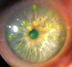
Filamentary keratitis is a condition in which strands (“filaments”) composed of degenerated epithelial cells and mucus develop on and adhere to the corneal surface causing pain and foreign body sensation.

The etiology of filamentary keratitis is related to an alteration in the components of the tear film and/or abnormalities of the corneal surface. It is associated with a number of ocular surface diseases and conditions.
Any alteration of the tear film or corneal surface can increase the risk of filamentary keratitis.
Common risk factors include:
- Aqueous tear deficiency as in keratoconjunctivitis sicca (or dry eye)
- Corneal exposure (e.g. seventh nerve palsy)
- Occlusion abnormalities such as blepharoptosis
- Ocular surgery (e.g. keratoplasty)
- Systemic diseases with effects on the ocular surface (e.g. Sjogren’s syndrome)
- Extended use of anticholinergic medications
- Superior limbic keratoconjunctivitis
- Other ocular surface abnormalities.
MANAGEMENT
General treatment
Filamentary keratitis treatment can be challenging and is often chronic. Paramount in the overall treatment of filamentary keratitis is the management of underlying conditions such as dry eye syndrome, medication toxicity, and blepharoptosis.
Medical therapy
First-line treatment includes topical therapy with lubricant drops and ointment. Low water-content bandage contact lenses may be helpful temporarily in cases that do not respond to lubrication alone.
The bandage contact lens should be used in combination with artificial tears and prophylactic topical antibiotics. A mucolytic agent such as 10% N-Acetylcysteine can be used topically to decrease the viscosity of the mucinous component of the tear film.
Topical sodium chloride drops may also help by deturgescing and compacting the corneal epithelium. Adjunct treatment with botulinum toxin may be considered in refractory cases. There are case reports and small case series that demonstrate the efficacy of sutureless amniotic membrane grafts to manage this condition.
Another possible treatment for refractory keratitis is autologous serum eye drops. Anti-inflammatory treatment will be needed, such as topical steroids (pulse or chronic), cyclosporine, and lifitegrast.
Medical follow up
Patients should be re-examined shortly after the initiation of medical therapy. Bandage contact lenses should be left in place for no more than one month.
Surgery
Filaments can be removed at the slit lamp using the jeweler’s forceps. Care should be taken to avoid disrupting the epithelium at the base of the filament if possible. Manual removal of the filaments may help in alleviating symptoms temporarily but is only a temporary measure and is not successful without concurrent medical treatment.
Punctal occlusion may also be helpful in cases of underlying aqueous tear deficiency either using temporary plugs or cauterization as a permanent solution.
HOW TO TAKE SLIT-LAMP EXAM IMAGES WITH A SMARTPHONE?
Smartphone slit-lamp photography is the new advancement in the field of science and technology in which photographs of the desired slit-lamp finding can be taken with smartphones by using the slit-lamp adapters.
Slit-lamp Smartphone photography
REFERENCES
- Davidson RS, Mannis MJ. Filamentary Keratitis. In: Krachmer JH, Mannis MJ, Holland EJ, editors. Cornea. Vol 1. 3rd ed. Philadelphia: Elsevier/Mosby; 2011. P. 1093-96.
- Van Meter WS, Katz D, Cook BG. Filamentary Keratitis. In: Holland EJ, Mannis MJ, Lee WB, editors. Ocular Surface Disease: Cornea, Conjunctiva, and Tear Film. Philadelphia: Elsevier Saunders; 2013. P. 213-16.
- Zaidman GW, Geeraets R, Paylor RR, et al. The histopathology of filamentary keratitis. Arch Ophthalmology 1985; 103: 1178-81.
- Tanioka H, Yokoi N, Komuro A, et al. Investigation of the corneal filament in filamentary keratitis. Invest Ophthalmol Vis Sci 2009; 50:3696-702.
- Gumus K, Lee S, Yen MT, Pflugfelder SC. Botulinum toxin injection for the management of refractory filamentary keratitis. Arch Ophthalmol. 2012 Apr;130(4):446-50. doi: 10.1001/archophthalmol.2011.2713. PMID: 22491914.
- Case report on biotissue website; Harthan JS, Sicks LA, “Sutureless Amniotic Membrane (Prokera) for Filamentary Keratitis: A Case Series, J Dry EyeOcc Surf Dis Vol 2(SP1):e10-e16; May 9, 2019.
