
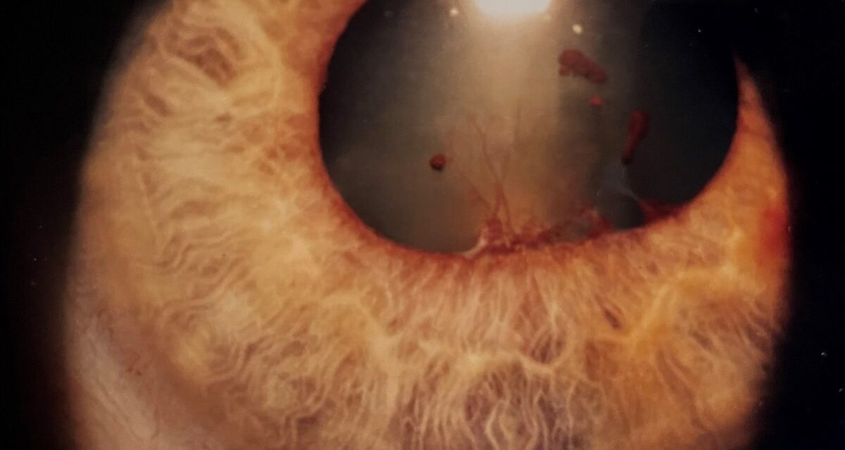
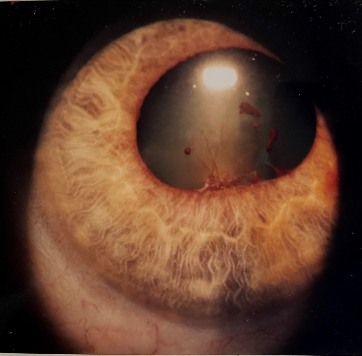
Posterior synechiae are adhesions between the posterior iris surface and the anterior lens capsule, formed when inflammatory fibrin and cellular debris in the anterior chamber organize into fixed bands during an episode of anterior uveitis.
They are one of the more consequential physical sequelae of intraocular inflammation, because unlike most other markers of uveitis activity, synechiae do not simply resolve once the inflammation is controlled — the adhesion, once formed, is usually permanent unless broken mechanically or pharmacologically early in its course.
Their clinical importance goes beyond cosmetic pupil irregularity: extensive synechiae can obstruct aqueous flow through the pupil and precipitate a specific, dangerous form of secondary glaucoma that requires urgent recognition and treatment.
Pathogenesis
During active anterior uveitis, breakdown of the blood-aqueous barrier allows fibrin, inflammatory cells, and protein to accumulate in the anterior chamber.
Where the posterior iris surface and anterior lens capsule are in close apposition — which is their entire circumference at rest — this exudate can organize into a fibrous adhesion within days if the pupil remains constricted and immobile during the inflammatory episode.
Chronic or recurrent uveitis produces the most extensive synechiae, because each flare adds the opportunity for new adhesions to form, if the pupil is not kept mobile with cycloplegic agents during active disease, gradually building on damage from earlier episodes rather than starting fresh each time.
This time-dependent nature of synechiae formation is precisely why cycloplegic treatment is started early and proactively during an acute uveitis flare, rather than reserved only for patients who have already begun to develop visible adhesions.
Clinical Findings
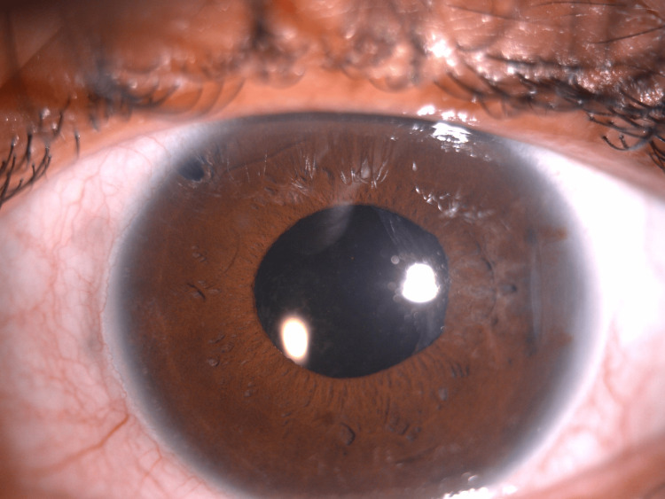
- Irregular, often festooned or scalloped pupil margin, rather than the normal round contour
- Poor or asymmetric pupillary dilation with mydriatic agents, since adherent segments of iris cannot move
- Pigment deposits on the anterior lens capsule at sites of prior adhesion, sometimes visible even after the synechia itself has been broken
- In extensive cases, complete adhesion around the entire pupil margin — seclusio pupillae — which prevents any aqueous flow from the posterior to anterior chamber
A distinctive irregular pupil in a patient with a history of red eye, even remote, should prompt specific questioning about prior uveitis, because the synechiae themselves can be the only remaining sign of an episode the patient may not have recognized as significant at the time, sometimes discovered only incidentally years later during an unrelated routine eye exam.
All-fit Slit-Lamp Adapter
Record and share exactly what you see at the slit lamp. One adapter fits any slit lamp or surgical microscope — and any smartphone.
From Choroida — the team behind this siteIris Bombe and Angle-Closure Glaucoma
When synechiae extend around the full circumference of the pupil (seclusio pupillae), aqueous produced by the ciliary body cannot pass from the posterior chamber to the anterior chamber through the normal pupillary route.
Pressure builds behind the iris, causing it to bow forward into a convex configuration — iris bombe — which pushes the peripheral iris against the trabecular meshwork and can precipitate acute angle-closure glaucoma, a genuine ophthalmic emergency in this context.
This is different in mechanism, though similar in urgency, to primary angle-closure glaucoma, and recognizing that the underlying cause is inflammatory rather than purely anatomic changes the immediate treatment approach, since addressing the inflammation itself is just as important as relieving the acute pressure elevation.
Differential Diagnosis of an Irregular Pupil
- Traumatic mydriasis or sphincter tear — history of blunt trauma, often with a peaked or scalloped margin from focal tears rather than adhesion
- Iris coloboma — congenital, typically inferonasal, present from birth
- Prior intraocular surgery with peripheral iridectomy or iridotomy
- Neovascular glaucoma with ectropion uveae — the pupil margin is pulled anteriorly by fibrovascular membrane rather than adhered posteriorly
- Adie’s tonic pupil — irregular, sluggish reaction, but without a history of intraocular inflammation and typically associated with light-near dissociation
Management and Prevention
Cycloplegic and mydriatic agents (cyclopentolate, homatropine, or atropine, depending on the severity and chronicity of inflammation) are used during active anterior uveitis specifically to keep the pupil mobile and prevent synechiae from forming or extending, which is one of the core reasons cycloplegia is standard therapy in uveitis, alongside its role in reducing ciliary spasm and pain.
Once an adhesion has formed, aggressive cycloplegia combined with topical corticosteroids in the early days can sometimes break a fresh synechia before it fully organizes, though established, longstanding adhesions are usually permanent, which is why prompt treatment at the very start of a flare offers the best chance of preventing permanent pupillary scarring.
Acute iris bombe with impending or established angle closure is treated urgently with laser peripheral iridotomy, or surgical iridectomy if laser access is not feasible, to create an alternative pathway for aqueous flow and relieve the pressure differential between chambers.
Longer-term, controlling the underlying uveitis to prevent further synechiae formation, and monitoring intraocular pressure closely in eyes with any degree of synechiae, are the mainstays of ongoing care, since even partial, incomplete synechiae can gradually worsen over subsequent flares if the pupil is not adequately protected during each episode.
Even after a synechia has become permanent, the goal of ongoing management shifts from breaking the adhesion to preventing further ones from forming, since additional synechiae in an eye already compromised by prior scarring further increases the cumulative risk of angle-closure glaucoma over time.


Document what you see
Two smartphone imaging tools built for everyday clinic use — one for the slit lamp, one for the fundus.
From Choroida — the team behind this siteReferences
- Jones NP. Glaucoma in uveitis. Eye.
- Moorthy RS, Mermoud A, Baerveldt G, et al. Glaucoma associated with uveitis. Survey of Ophthalmology.
- American Academy of Ophthalmology. Basic and Clinical Science Course, Section 9: Intraocular Inflammation and Uveitis.
- Sallam A, Sheth HG, Habot-Wilner Z, Lightman S. Outcome of raised intraocular pressure in uveitic eyes with and without a corticosteroid-induced hypertensive response. American Journal of Ophthalmology.
