
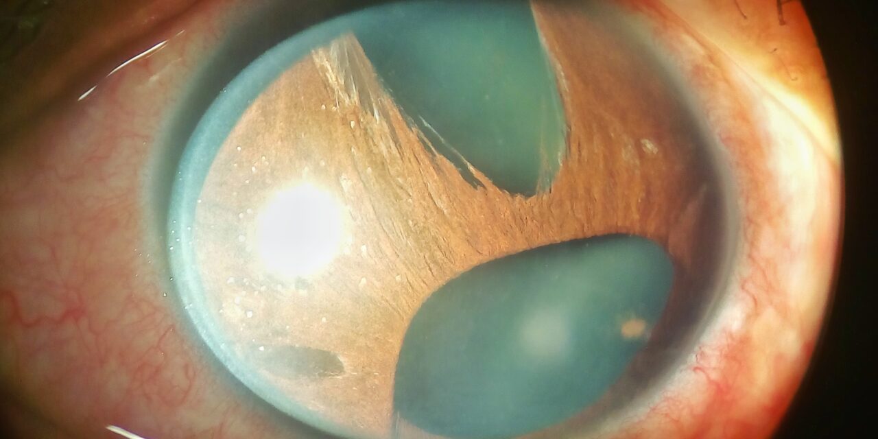
Iridocorneal Endothelial Syndrome is a disease in which the cornea’s innermost cells forget their boundaries.
The corneal endothelium proliferates and creeps across the angle and onto the iris, dragging it out of shape and sealing the drainage angle shut.
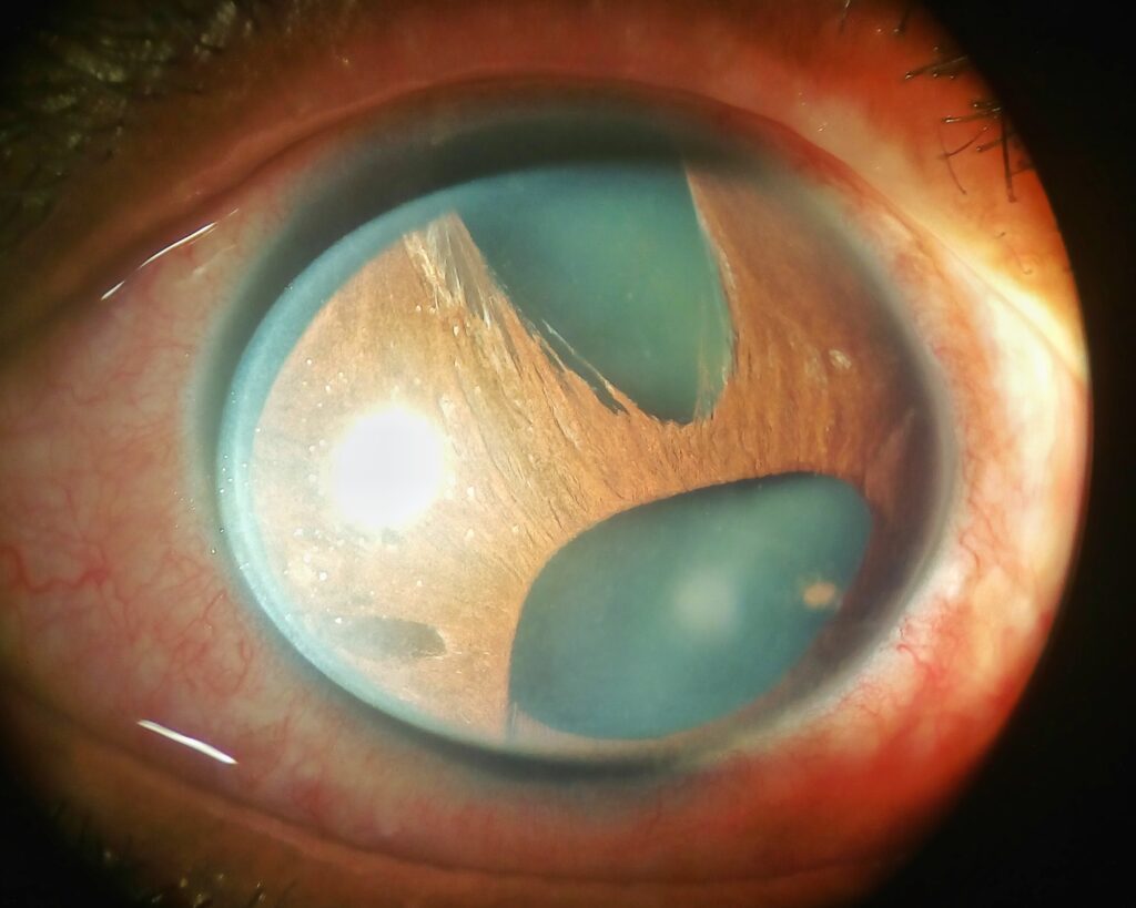
The result is a strikingly distorted iris, a swollen cornea, and a stubborn secondary glaucoma, almost always in one eye of a middle-aged woman.
For clinicians, recognizing Iridocorneal Endothelial Syndrome (ICE) matters because it is a unilateral, progressive cause of glaucoma and corneal decompensation that behaves quite differently from ordinary open-angle disease.
An asymmetric iris, a hammered-silver endothelium, and a high pressure in one eye are the clues that bring it to mind.
What Is Iridocorneal Endothelial Syndrome?
ICE syndrome is a spectrum of disorders in which an abnormal corneal endothelium migrates across the anterior chamber angle and iris surface.
It is traditionally divided into three overlapping variants:
- Chandler syndrome — dominated by a beaten-metal endothelium and corneal oedema
- Essential (progressive) iris atrophy — dominated by iris thinning, holes, and displacement
- Cogan-Reese syndrome — dominated by pigmented pedunculated iris nodules
All three share the same underlying problem: a membrane of abnormal endothelial cells that contracts over the angle and iris.
Epidemiology and Cause
The demographic pattern is fairly consistent.
- Typically affects middle-aged adults, more often women
- Almost always unilateral and non-familial
- A viral trigger, particularly herpes simplex, has been implicated in the abnormal endothelium
The unilateral, sporadic nature helps separate ICE from the inherited endothelial dystrophies.
All-fit Slit-Lamp Adapter
Record and share exactly what you see at the slit lamp. One adapter fits any slit lamp or surgical microscope — and any smartphone.
From Choroida — the team behind this siteClinical Presentation
Symptoms
- Blurred vision from corneal oedema, often worse in the morning
- A change in the appearance of the pupil or iris, sometimes noticed by the patient
- Symptoms of raised pressure when glaucoma is advanced
Examination Findings
- A beaten-bronze or hammered-silver endothelium on the affected cornea
- Corneal oedema, especially in the Chandler variant
- Iris changes — corectopia, polycoria, atrophy, and pedunculated nodules
- Broad peripheral anterior synechiae that close the angle on gonioscopy
A distorted, off-centre pupil with peripheral anterior synechiae in one eye is highly suggestive of ICE.
Diagnostic Evaluation
Endothelial imaging and gonioscopy confirm the diagnosis.
- Specular or confocal microscopy shows characteristic abnormal endothelial cells, the so-called ICE cells
- Gonioscopy demonstrates peripheral anterior synechiae and angle closure
- Slit-lamp examination documents the corneal and iris changes
- Intraocular pressure and optic nerve assessment stage the glaucoma
The abnormal endothelium seen on specular microscopy is the finding that anchors the diagnosis.
Differential Diagnosis
A few conditions produce overlapping features:
- Posterior polymorphous corneal dystrophy — bilateral and inherited
- Fuchs endothelial dystrophy — bilateral, without iris distortion or synechiae
- Axenfeld-Rieger syndrome — bilateral, congenital, and often familial
- Posterior polymorphous and iridocorneal overlap can be resolved with laterality and specular microscopy
Unilaterality, acquired onset, and the ICE cells distinguish it from these bilateral, inherited conditions.
Management
Treatment targets the two threats: pressure and cornea.
- Glaucoma is managed medically first, but often progresses to surgery; glaucoma drainage devices tend to outperform trabeculectomy because the membrane can occlude a trabeculectomy
- Corneal decompensation may require endothelial or penetrating keratoplasty
- Laser trabeculoplasty is generally ineffective because the angle is covered by membrane
Because the abnormal endothelium keeps advancing, management is about controlling its consequences rather than curing it.
Prognosis
The course is slowly progressive.
- Glaucoma can be difficult to control and is the main threat to vision
- Corneal decompensation may develop over time
- Early, structured management preserves useful vision in most eyes
In practice, the value of recognising ICE is anticipating its glaucoma and corneal complications rather than being surprised by them.
Would you like to document corneal and iris findings with your smartphone?


Document what you see
Two smartphone imaging tools built for everyday clinic use — one for the slit lamp, one for the fundus.
From Choroida — the team behind this siteReferences
- Shields MB. “Progressive Essential Iris Atrophy, Chandler’s Syndrome, and the Iris Nevus (Cogan-Reese) Syndrome: A Spectrum of Disease.” Survey of Ophthalmology. 1979;24(1):3–20.
- Sacchetti M, Mantelli F, Marenco M, et al. “Diagnosis and Management of Iridocorneal Endothelial Syndrome.” BioMed Research International. 2015;2015:763093.
- Alvarado JA, Underwood JL, Green WR, et al. “Detection of Herpes Simplex Viral DNA in the Iridocorneal Endothelial Syndrome.” Archives of Ophthalmology. 1994;112(12):1601–1609.
- American Academy of Ophthalmology. “Iridocorneal Endothelial Syndrome.” EyeWiki.
Test yourself
A few questions straight from this article.
-
What is the core abnormality shared by all variants of iridocorneal endothelial (ICE) syndrome?
In ICE syndrome an abnormal corneal endothelium migrates across the anterior chamber angle and iris surface, forming a membrane that contracts and distorts both. -
Which variant of ICE syndrome is dominated by pigmented pedunculated iris nodules?
Cogan-Reese syndrome is the ICE variant characterised by pigmented pedunculated iris nodules, while Chandler is dominated by corneal oedema and essential iris atrophy by iris holes. -
Which demographic and laterality pattern is typical of ICE syndrome?
ICE syndrome typically affects middle-aged adults, more often women, and is almost always unilateral and non-familial, which separates it from the inherited endothelial dystrophies. -
Which infectious agent has been implicated in the abnormal endothelium of ICE syndrome?
A viral trigger, particularly herpes simplex, has been implicated in producing the abnormal corneal endothelium of ICE syndrome. -
What is the classic slit-lamp appearance of the corneal endothelium in ICE syndrome?
The affected cornea shows a beaten-bronze or hammered-silver endothelium, often with corneal oedema that is most marked in the Chandler variant. -
What does gonioscopy characteristically demonstrate in ICE syndrome?
The contracting endothelial membrane draws the iris forward, producing broad peripheral anterior synechiae that close the angle on gonioscopy. -
Which investigation anchors the diagnosis of ICE syndrome by showing the characteristic ICE cells?
Specular or confocal microscopy shows the characteristic abnormal endothelial cells known as ICE cells, and this is the finding that anchors the diagnosis. -
Which features separate Fuchs endothelial dystrophy from ICE syndrome?
Fuchs endothelial dystrophy is bilateral and produces no iris distortion or synechiae, whereas ICE syndrome is unilateral, acquired and remodels the iris and angle. -
Which glaucoma operation tends to perform better in ICE syndrome, and why?
Glaucoma drainage devices tend to outperform trabeculectomy in ICE syndrome because the advancing endothelial membrane can occlude a trabeculectomy; laser trabeculoplasty is generally ineffective as the angle is covered by membrane. -
What is the main threat to vision over the long course of ICE syndrome?
The course is slowly progressive, and the glaucoma can be difficult to control and is the main threat to vision, with corneal decompensation developing over time.
