
Internuclear ophthalmoplegia (INO) is impaired adduction of one eye on horizontal gaze, accompanied by nystagmus of the abducting fellow eye, caused by a lesion of the medial longitudinal fasciculus.
It is a small, precise finding that punches well above its size diagnostically: in a younger patient it points toward multiple sclerosis, and in an older patient it points toward brainstem stroke, and distinguishing between the two on clinical grounds alone is often possible before any imaging is done.
Anatomy and Pathophysiology
The medial longitudinal fasciculus is a heavily myelinated tract that connects the abducens nucleus on one side to the contralateral oculomotor nucleus, specifically the subnucleus supplying the medial rectus.
It is the anatomical basis for conjugate horizontal gaze: when the abducens nucleus fires to abduct one eye, the signal travels via the MLF to drive simultaneous adduction of the fellow eye, so the two eyes move together rather than independently.
A lesion of the MLF interrupts this crossed signal. On attempted gaze away from the side of the lesion, the ipsilateral eye fails to adduct normally, while the contralateral eye abducts but shows nystagmus, generally attributed to an attempt by the central nervous system to overdrive the intact abducens pathway to compensate for the weak adduction on the other side.
Convergence is typically preserved in INO, because the pathway mediating convergence bypasses the MLF and uses a different route. When convergence is also impaired alongside a failure of adduction, this points toward a different, usually more anterior or mesencephalic, lesion rather than a pure MLF problem, and should prompt reconsideration of the localisation.
Clinical Presentation
Patients typically report horizontal diplopia, worse on gaze away from the side of the lesion, and sometimes oscillopsia related to the nystagmus.
Bilateral INO can also produce vertical diplopia or a sense of unsteady, jumping vision, and patients do not always describe the deficit in a way that immediately suggests a gaze-direction pattern, which makes the eye movement examination the key step rather than the history alone.
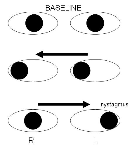
On examination, look for slowed or incomplete adduction of the affected eye on gaze away from the lesion side, with the fellow, abducting eye showing nystagmus in the direction of gaze.
In milder or subtler cases, the adduction deficit can be easy to miss on casual observation, and formal saccadic velocity testing, timing how quickly the eye reaches its adducted target, will often reveal slowing that is not obvious to the naked eye.
Convergence should specifically be tested and is typically intact. Vertical gaze and other cranial nerve function should be assessed as part of a full localising examination, since additional findings can help pin down the lesion more precisely within the brainstem.
Bilateral INO, with adduction deficits on gaze to both sides, is a particularly useful localising finding because it strongly suggests a single midline process, most classically multiple sclerosis, rather than a discrete unilateral vascular lesion.
All-fit Slit-Lamp Adapter
Record and share exactly what you see at the slit lamp. One adapter fits any slit lamp or surgical microscope — and any smartphone.
From Choroida — the team behind this siteAetiology
In younger patients, especially with bilateral involvement, demyelination from multiple sclerosis is the leading cause, and INO can be one of the presenting signs of MS rather than a late complication.
In older patients, particularly with unilateral INO and vascular risk factors, brainstem infarction affecting the MLF is the more likely cause. Other, less common causes include brainstem tumour, infection or abscess, and trauma.
As a general pattern, bilateral INO in a younger person raises suspicion for demyelination, while unilateral INO in an older person with vascular risk factors raises suspicion for stroke, though this is a guide rather than a rule and both age groups can, less commonly, have either cause.
Differential Diagnosis
- Medial rectus palsy from a third nerve lesion, which typically comes with other oculomotor findings such as ptosis or pupil involvement, unlike isolated INO
- Myasthenia gravis, which can mimic almost any pattern of ophthalmoplegia, including one resembling INO, and should be considered particularly when the pattern is variable or accompanied by fatigable ptosis
- Thyroid eye disease with restrictive myopathy, distinguished by a positive forced duction test and the absence of nystagmus in the fellow eye
- Wernicke encephalopathy and other brainstem processes affecting conjugate gaze more diffusely, usually with additional systemic or neurological findings
A forced duction test is a useful discriminator when the diagnosis is unclear: a genuinely restrictive process, like thyroid eye disease, will show resistance to passive movement, whereas INO, being neurogenic, will not.
Diagnostic Evaluation
MRI of the brain, with particular attention to the brainstem, is the key investigation and will usually identify a demyelinating plaque or an area of infarction corresponding to the clinical findings.
In a younger patient with findings suggesting demyelination, further workup for multiple sclerosis is appropriate, including MRI of the spinal cord where clinically indicated and consideration of cerebrospinal fluid analysis.
In an older patient with a vascular pattern, standard stroke workup applies: vascular imaging, cardiac evaluation, and assessment of modifiable vascular risk factors.
Management
Management addresses the underlying cause rather than the eye movement finding itself. In multiple sclerosis, this means disease-modifying therapy and, for an acute relapse, corticosteroids, managed jointly with neurology.
In stroke, it means standard acute stroke management and secondary prevention.
Diplopia can be managed symptomatically with prism correction or monocular occlusion while the underlying process is treated or while spontaneous recovery occurs, and referral to neurology is appropriate for any newly diagnosed INO given the range of underlying causes and the need for directed further workup.
Prognosis
Vascular INO frequently improves over weeks to months as the surrounding tissue recovers, though some patients are left with a mild, persistent deficit.
INO related to multiple sclerosis can improve with treatment of the underlying relapse but has a greater tendency to recur, either as the same finding or as a new lesion elsewhere along the pathway, reflecting the relapsing nature of the underlying disease.
Functional outcome is generally good regardless of cause, since the deficit itself, even when it persists, is often mild enough that patients adapt well, particularly once any diplopia has been addressed with prisms or occlusion.


Document what you see
Two smartphone imaging tools built for everyday clinic use — one for the slit lamp, one for the fundus.
From Choroida — the team behind this siteReferences
- Leigh RJ, Zee DS. The Neurology of Eye Movements. Oxford University Press.
- Frohman EM, Frohman TC, Zee DS, et al. The neuro-ophthalmology of multiple sclerosis. The Lancet Neurology. 2005.
- Keane JR. Internuclear ophthalmoplegia: unusual causes in 114 of 410 patients. Archives of Neurology. 2005.
- Internuclear Ophthalmoplegia. EyeWiki, American Academy of Ophthalmology.
- Internuclear Ophthalmoplegia. StatPearls, NCBI Bookshelf.
Test yourself
A few questions straight from this article.
-
Which combination of findings defines internuclear ophthalmoplegia?
INO is impaired adduction of one eye on horizontal gaze, with nystagmus in the abducting fellow eye. The responsible lesion sits in the medial longitudinal fasciculus. -
The medial longitudinal fasciculus connects the abducens nucleus to which structure?
The MLF links the abducens nucleus to the contralateral oculomotor nucleus, specifically the medial rectus subnucleus. This crossed connection is the anatomical basis of conjugate horizontal gaze. -
In a unilateral internuclear ophthalmoplegia, when does the adduction deficit appear?
On gaze away from the lesion side, the ipsilateral eye fails to adduct normally while the contralateral eye abducts with nystagmus. The nystagmus is attributed to central overdrive of the intact abducens pathway. -
What does preserved convergence indicate in a patient with an adduction deficit?
Convergence bypasses the MLF by a different route, so it is typically preserved in INO. Adduction failure combined with impaired convergence suggests a more anterior or mesencephalic lesion and should prompt reconsidering the localisation. -
What does bilateral internuclear ophthalmoplegia most strongly suggest?
Adduction deficits on gaze to both sides point to one midline process rather than a discrete unilateral vascular lesion. Demyelination from multiple sclerosis is the classic cause, and INO can be a presenting sign of MS. -
Which cause is most likely for unilateral internuclear ophthalmoplegia in an older patient with vascular risk factors?
Unilateral INO in an older patient with vascular risk factors most often reflects brainstem infarction. This is a guide rather than a rule, since either age group can less commonly have the other cause. -
How does a forced duction test help separate internuclear ophthalmoplegia from thyroid eye disease?
A genuinely restrictive process such as thyroid eye disease resists passive movement, whereas INO is neurogenic and offers no resistance. Thyroid restriction also lacks nystagmus in the fellow eye. -
Which investigation is the key study in a newly diagnosed internuclear ophthalmoplegia?
Brain MRI focused on the brainstem usually identifies a demyelinating plaque or an area of infarction matching the clinical findings. Further workup then follows the demyelinating or vascular pattern suggested. -
What is the principle guiding management of internuclear ophthalmoplegia?
Management addresses the underlying disease rather than the eye movement itself, with prisms or monocular occlusion used symptomatically. Neurology referral is appropriate for any newly diagnosed INO. -
How does the course of vascular internuclear ophthalmoplegia typically compare with that of multiple sclerosis?
Vascular INO frequently improves over weeks to months as surrounding tissue recovers, sometimes leaving a mild persistent deficit. INO from multiple sclerosis has a greater tendency to recur, reflecting the relapsing disease.

Writes and reviews the clinical articles on Educate. Every post is checked against peer-reviewed sources before it goes live.


