
![[Answered] What is Pinguecula & how to treat it ?](https://educate.choroida.com/wp-content/uploads/2022/09/pinguecula-3.LRG_-1280x640.jpg)
Case Report of Pinguecula
A 36-year-old man was referred to a clinic with an elevated yellowish mass on his conjunctiva in his left eye

which was associated with dry eye symptoms (i.e., soreness, scratchiness, dryness, grittiness, and burning)
and general discomfort, such as “tightening sensation” and “stiffness”.
Interestingly,
he reported having chronic inflammation of the aforementioned area for 2–3 years, for which he received topical lubricants and/or steroids, with temporary or partial improvement of ocular irritation.
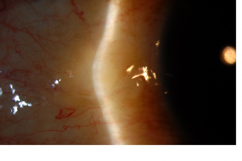
On clinical assessment, the left eye revealed a yellow-white, circular bump on the nasal conjunctiva adjacent to the limbus, mild/focal conjunctival injection, a fluorescein break-up time (FBUT) of 3.5 s, an abnormal fluorescein staining, and a Schirmer I test of 5 mm.
Optical coherence tomography (OCT) imaging evidenced a height at the most elevated point of the pinguecula of 740 µm.
Disease “Pinguecula”
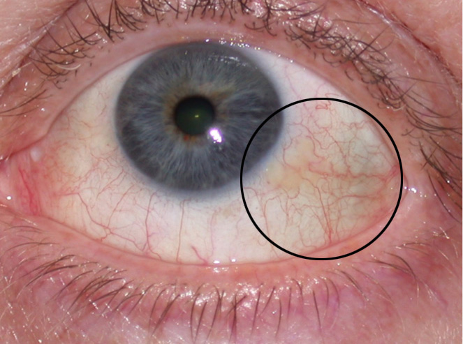
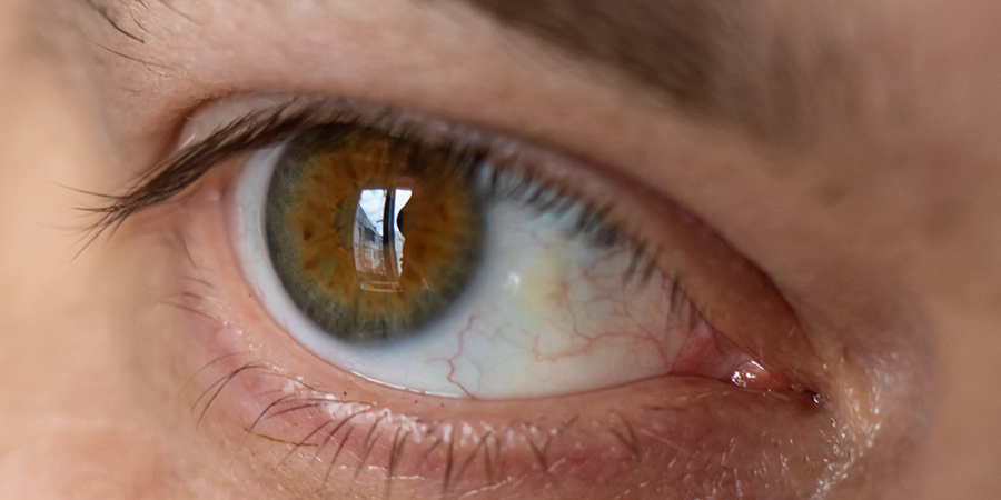
A relatively common non–malignant raised yellow-white lesion of the interpalpebral bulbar conjunctiva that does not involve the cornea and represents elastoic degeneration of subepithelial collagen with hyalinized connective tissue.
These fleshy lesions are typically found bilaterally and adjacent to the limbus of the nasal bulbar conjunctiva although they can be present temporally as well.
Pingueculae are thought to arise as a result of the effects of environmental irritants such as wind and dust and are associated with UV-light exposure and aging.
However, the evidence of the association between UV-light exposure and pingueculae remains limited.
It is thought that Pinguecula arises more commonly on the nasal side because light passing medially through the cornea focuses on the area of the nasal limbus
while the shadow of the nose reduces the intensity of light transmitted to the area of the temporal limbus.
Diagnosis of Pinguecula
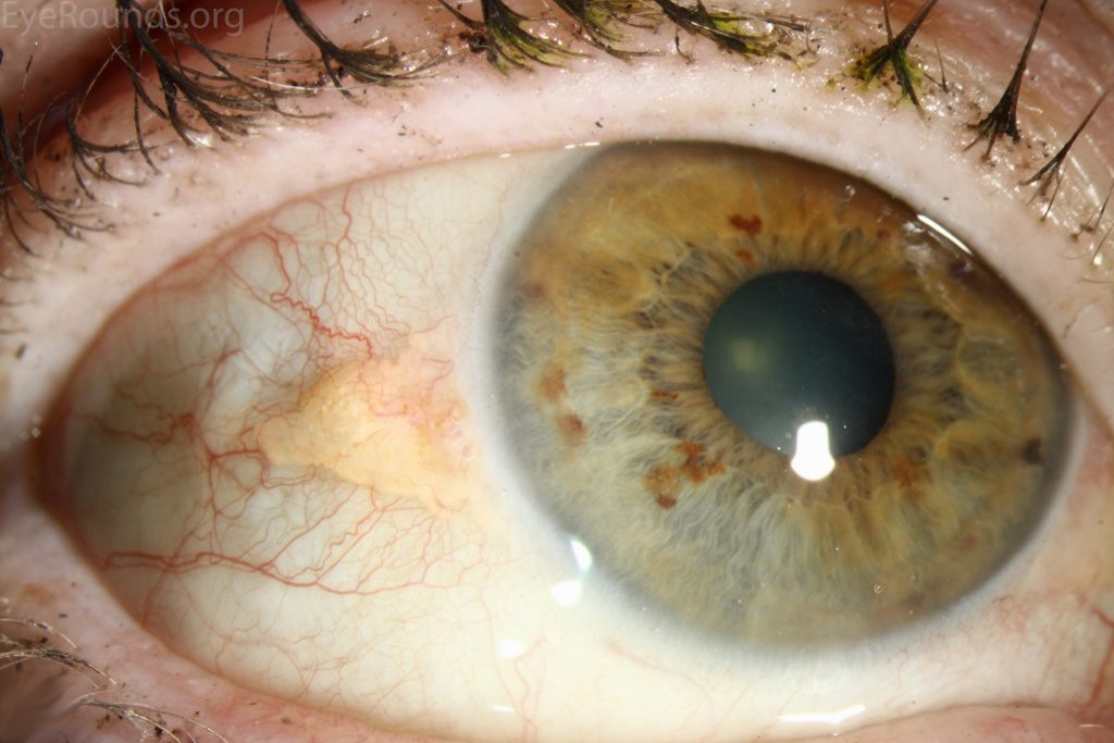
The diagnosis is usually made clinically by Slit-lamp biomicroscopy. The classic growth is raised and yellow-white in color, found in the interpalpebral fissure, and is more common nasally than temporally.
The lesion may be highly vascularized and injected or associated with punctuating epithelial erosions or Dellen (thinning of the adjacent cornea due to drying).
Management of Pinguecula
Lubrication with artificial tears and ointment can help with ocular surface irritation.
Excision is indicated only when pingueculae are cosmetically unacceptable or when they become chronically inflamed or interfere with successful contact lens wear.
Long-term use of topical steroid therapy should be discouraged due to adverse side effects but can but used judiciously in patients with inflamed pingueculae termed “pingueculitis”.
Topical indomethicin has also been demonstrated at reducing symptoms of inflammation.
Surgery:
Laser photocoagulation and surgical excision of pingueculae have both been used successfully with similar cosmetic outcomes.
A study of 30 patients showed surgical excision of symptomatic pinguecula with conjunctival autograft secured with fibrin glue improved not only cosmesis but also improved dry eye syndrome.
HOW TO TAKE SLIT-LAMP EXAM IMAGES WITH A SMARTPHONE?
Smartphone slit-lamp photography is the new advancement in the field of science and technology in which photographs of the desired slit-lamp finding can be taken with smartphones by using the slit-lamp adapters.
Slit-lamp Smartphone photography
REFERENCES
- Weisenthal, Robert W, Afshari, Natalie A, Bouchard, Charles S, Colby, Kathyrn A, Rootman, David S, Tu, Elmer Y, 2013, External Disease and Cornea, AAO, BCSC, Chapter 12, Depositions and Degenerations.
- Yam JC, Kwok AK. Ultraviolet light and ocular diseases. Int Ophthalmol. 2013 May: 14(2)187-94
- Mimura T, Obata H, Usui T, Mori M, Yamagami S, Funatsu H, Noma H, Amano S, Pinguecula and diabetes mellitus. Cornea. 2012 Mar;31(3):264-8.
- Dushku N, Reid TW, P53 expression in altered limbal basal cells of pingueculae, pterygia, and limbal tumors. Curr Eye Res. 1997 Dec;16(12):1179-92
- Machemer, Robert. Pterygium, Atlas of ophthalmology.com
- Bergmanson JP, Söderberg PG, The significance of ultraviolet radiation for eye diseases. A review with comments on the efficacy of UV-blocking contact lenses. Ophthalmic Physiol Opt. 1995 Mar;15(2):83-91
Slit-lamp Smartphone photography
