
Interstitial keratitis is inflammation of the corneal stroma without primary involvement of the epithelium or endothelium, and its classic cause, congenital syphilis, has become rare enough in many settings that the diagnosis is easy to miss the first time a clinician actually encounters it.
The disease itself is really an immune reaction to organisms or antigens that reached the cornea years earlier, which is why the presentation, often in an older child or young adult, can seem disconnected from any history the patient or family readily volunteers.
Pathophysiology
Stromal inflammation in interstitial keratitis is generally an immune-mediated response to an organism, most classically Treponema pallidum transmitted transplacentally in congenital syphilis, rather than active infection of the cornea itself at the time symptoms appear.
Vascularisation of the stroma follows, with vessels growing in from the limbus, and this vascular infiltration, together with stromal oedema and cellular infiltrate, is what produces the characteristic diffuse corneal haze.
Because the trigger was often present from infancy while the clinical disease manifests later, patients with congenital syphilitic interstitial keratitis frequently present in the first two decades of life, sometimes with no known history of syphilis in the family at all, since maternal infection may never have been diagnosed or disclosed.
Causes
- Congenital syphilis – the classic and historically most common cause, typically presenting between five and twenty years of age, often bilateral though not necessarily simultaneous in onset
- Acquired syphilis, less commonly associated with interstitial keratitis than the congenital form
- Herpes simplex and herpes zoster virus, producing a stromal keratitis that can closely resemble the syphilitic pattern
- Tuberculosis, Lyme disease, and other less common infectious associations
- Cogan syndrome, a rare autoimmune condition combining non-syphilitic interstitial keratitis with audiovestibular symptoms resembling Ménière disease, distinct from an infectious cause entirely
All-fit Slit-Lamp Adapter
Record and share exactly what you see at the slit lamp. One adapter fits any slit lamp or surgical microscope — and any smartphone.
From Choroida — the team behind this siteClinical Presentation
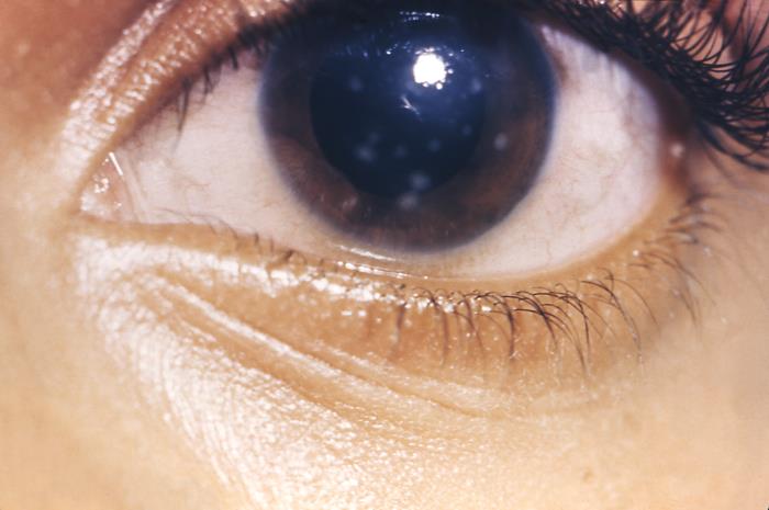
Onset is typically insidious, with photophobia, tearing, and gradually blurred vision developing over days to weeks, often in an older child or young adult with congenital syphilis, and frequently without pain proportional to the degree of inflammation visible on examination.
The cornea shows diffuse or patchy stromal haze with deep vascularisation, sometimes producing a pink or salmon-coloured appearance in more active disease from the combination of vessels and inflammatory infiltrate. As the acute inflammation resolves, the vessels can regress, leaving behind faint, empty “ghost vessels” that remain visible indefinitely and are a useful retrospective clue to prior interstitial keratitis even decades later.
Bilateral involvement is common in congenital syphilis, though the two eyes are frequently affected sequentially rather than simultaneously, sometimes separated by months to years.
Associated Systemic Findings
In congenital syphilis, interstitial keratitis is one component of Hutchinson’s triad, alongside Hutchinson teeth (notched, peg-shaped upper central incisors) and sensorineural hearing loss, and the presence of any one of these should prompt a search for the others.
Other stigmata of congenital syphilis, including saddle nose deformity and frontal bossing, may be present and support the diagnosis when found alongside the corneal findings.
Diagnostic Evaluation
Serological testing for syphilis, including both a non-treponemal test and a confirmatory treponemal-specific test, is essential in any patient presenting with a pattern suggestive of interstitial keratitis, since active disease at the time of ocular presentation is unusual and treponemal-specific tests remain positive lifelong regardless of prior treatment.
Audiometry should be arranged to assess for the sensorineural hearing loss component of Hutchinson’s triad, and dental examination can reveal the characteristic notched incisors when present.
Where herpetic keratitis is suspected instead, viral PCR or a trial of antiviral therapy can help distinguish it, and where Cogan syndrome is suspected, audiovestibular assessment and a broader autoimmune workup are appropriate given its different treatment implications.
Differential Diagnosis
- Herpetic stromal keratitis, which can closely mimic syphilitic interstitial keratitis but is typically unilateral and associated with a history of prior epithelial herpetic disease
- Cogan syndrome, distinguished by negative syphilis serology and associated audiovestibular symptoms
- Other causes of corneal vascularisation, including chronic contact lens wear or ocular surface disease, generally distinguished by a very different clinical context and history
- Acanthamoeba keratitis, typically more acutely painful with a ring infiltrate rather than the diffuse haze of interstitial keratitis
Management
Treating the Underlying Cause
Systemic treatment for confirmed syphilis, generally penicillin following current guidelines, is essential, both to address any residual active infection and as part of comprehensive management of the underlying congenital syphilis, coordinated with paediatric infectious disease specialists.
Controlling Ocular Inflammation
Topical corticosteroids are the mainstay of treatment for the acute inflammatory component, often needed for a prolonged course with gradual tapering, alongside cycloplegics for comfort and to reduce the risk of posterior synechiae during active inflammation.
Herpetic Interstitial Keratitis
When herpes simplex or zoster is the identified cause, antiviral therapy is added alongside corticosteroids, following the same principles used for herpetic stromal keratitis more broadly.
Prognosis
Visual outcome depends on the severity and duration of stromal scarring left behind once active inflammation resolves; many patients retain reasonably good vision, though dense central scarring or significant residual vascularisation can cause permanent visual impairment.
Ghost vessels typically persist indefinitely even after successful treatment, though they generally do not by themselves cause significant visual symptoms once the active inflammatory phase has resolved.
Recurrence is uncommon once the underlying infection has been adequately treated, though the corneal changes and any associated hearing loss from congenital syphilis are permanent, which is why the ophthalmic diagnosis often becomes the entry point for a fuller assessment and treatment of the underlying systemic disease.


Document what you see
Two smartphone imaging tools built for everyday clinic use — one for the slit lamp, one for the fundus.
From Choroida — the team behind this siteReferences
- Chao J, Khurana R. Ophthalmic manifestations of syphilis. Cornea. 2011.
- Wilhelmus KR, Grierson I, Watson PG. Interstitial keratitis. Cornea. 1981.
- Grasland A, Pouchot J, Hachulla E, et al. Typical and atypical Cogan’s syndrome. Rheumatology. 2004.
- Interstitial Keratitis. EyeWiki, American Academy of Ophthalmology.
- Congenital Syphilis. StatPearls, NCBI Bookshelf.
Test yourself
A few questions straight from this article.
-
Which part of the cornea is primarily inflamed in interstitial keratitis?
Interstitial keratitis is defined as inflammation of the corneal stroma without primary involvement of the epithelium or endothelium. -
What drives the stromal inflammation of interstitial keratitis in congenital syphilis?
The disease is an immune-mediated response to organisms or antigens that reached the cornea earlier, rather than active corneal infection at the time symptoms appear. -
At what age does interstitial keratitis from congenital syphilis typically present?
Although the trigger is present from infancy, clinical disease usually manifests later, most often in the first two decades of life. -
What corneal appearance characterises actively inflamed interstitial keratitis?
Stromal oedema, cellular infiltrate and deep vessels growing in from the limbus combine to give a pink or salmon-coloured appearance in active disease. -
Which corneal finding remains as a lifelong clue to previous interstitial keratitis?
Once inflammation settles the stromal vessels regress but leave empty ghost vessels that stay visible indefinitely, a useful retrospective clue decades later. -
Interstitial keratitis forms part of Hutchinson's triad alongside which two findings?
Hutchinson's triad comprises interstitial keratitis, notched peg-shaped upper central incisors, and sensorineural hearing loss; finding one should prompt a search for the others. -
Which investigation is essential in a patient presenting with interstitial keratitis?
Serological testing for syphilis using both a non-treponemal test and a confirmatory treponemal-specific test is essential in any suggestive corneal presentation. -
What typically distinguishes herpetic stromal keratitis from syphilitic interstitial keratitis?
Herpetic stromal keratitis can look very similar but is usually unilateral and accompanied by a history of earlier epithelial herpetic disease. -
What is the mainstay of treating the acute ocular inflammation in interstitial keratitis?
Topical steroids control the stromal inflammation and often need a prolonged, gradually tapered course, with cycloplegics added for comfort and to reduce posterior synechiae. -
What most determines the final visual outcome in interstitial keratitis?
Many patients retain reasonable vision, but dense central scarring or significant residual vascularisation left after the inflammation resolves can cause permanent impairment.

Writes and reviews the clinical articles on Educate. Every post is checked against peer-reviewed sources before it goes live.


