
CASE REPORT
A 49-year-old man with a history of Type II Diabetes and recent ocular trauma. He presented with decreased visual acuity and pain in his right eye (RE). Upon examination, the RE showed improved visual acuity with correction, but high intraocular pressure and signs of inflammation such as corneal edema and conjunctival hyperemia.
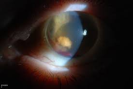
The vitreous appeared cloudy, hindering a detailed view of the optic nerve and macula. The contralateral eye appeared normal.
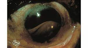
Further investigations were conducted, including B-mode ultrasound, which revealed increased echogenicity suggestive of vitreous hemorrhage.
A paracentesis procedure was performed to obtain a sample of aqueous humor, which was stained with hematoxylin-eosin, confirming the presence of spherical erythrocytes containing degenerated hemoglobin (Heinz bodies).
Additionally, anterior segment optical coherence tomography (OCT) showed moderate cellularity in the anterior chamber. Based on these clinical findings, the patient was diagnosed with ghost cell glaucoma (GCF).
Ghost Cell Glaucoma DISEASE entity
Ghost cell glaucoma is a secondary open-angle glaucoma caused by degenerated red blood cells (ghost cells) obstructing the trabecular meshwork.
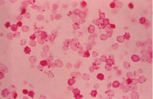
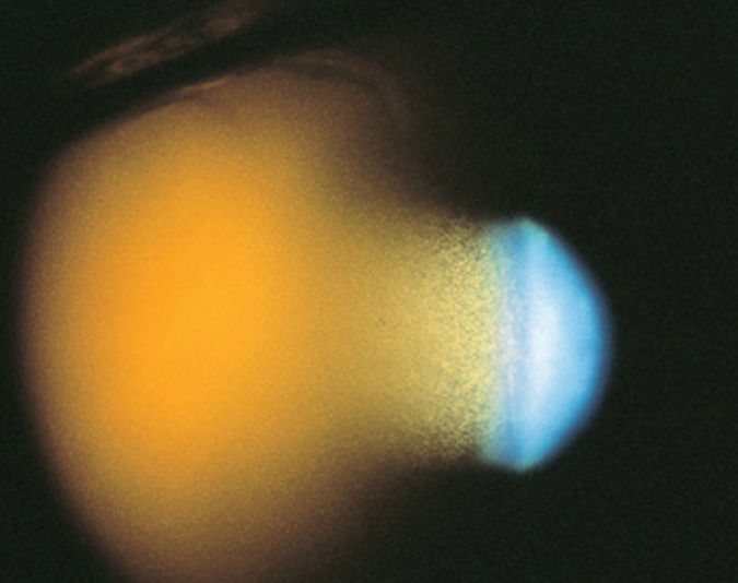
Following vitreous hemorrhage, blood breakdown products may accumulate in the anterior chamber (AC). Hemolyzed erythrocytes obstruct aqueous outflow through the trabecular meshwork leading to secondary open-angle glaucoma.
Ghost cell glaucoma is closely associated with vitreous hemorrhage. Causes of ghost cell glaucoma include causes of vitreous hemorrhage such as ocular trauma, systemic diseases such as diabetes or sickle cell disease/trait, iritis (Fuchs heterochromic iridocyclitis, herpes simplex, herpes zoster, etc.), intraocular tumors (retinoblastoma, malignant melanoma, etc.), uveitis glaucoma hyphema syndrome, rubeosis iridis, iris varices, papillary micro hemangiomas, and ocular surgery including but not limited to cataract extraction, laser trabeculoplasty, and iridotomy.
One case report of ghost cell glaucoma after a snake bite has been reported. Ghost cell glaucoma can also be associated with spontaneous ocular hemorrhages.
The symptoms relate to the etiology. Pain secondary to trauma or surgery may be experienced by the patient. However, less pain is often reported than expected from a severely elevated IOP. Patients with high IOP may also present with blurry vision, headache, brow ache, nausea, and/or vomiting.
All-fit Slit-Lamp Adapter
Record and share exactly what you see at the slit lamp. One adapter fits any slit lamp or surgical microscope — and any smartphone.
From Choroida — the team behind this siteGhost Cell Glaucoma MANAGEMENT
Ghost cell glaucoma usually resolves once the vitreous hemorrhage has cleared. Medical therapy with aqueous suppressants is the preferred initial approach.
Surgical intervention is often necessary because of persistently elevated intraocular pressure despite maximum medical therapy.
Medical Therapy
Aqueous suppressants are the first-line approach. Monotherapy or a combination of topical alpha-adrenergic agonists, beta-adrenergic blockers, parasympathomimetics, prostaglandin analogs, and carbonic anhydrase inhibitors may be used.
An oral carbonic anhydrase inhibitor may be added. Intravenous Mannitol or Diamox may be used for extremely high IOPs in acute settings. This may occur once cells re-accumulate in the trabecular meshwork after initial clearing.
Surgery
Surgery might be required to remove the ghost cells from the AC and decrease the stress on the trabecular meshwork. This can be accomplished by AC paracentesis and irrigation, pars plana vitrectomy (PPV), and/or a trabeculectomy.
If AC washout successfully lowers the intraocular pressure but the pressure rises again secondary to the reaccumulation of ghost cells originating in the vitreous, a washout of the AC can be repeated. If this relatively simple and safe procedure is unsuccessful, a vitrectomy to remove the contents of the vitreous cavity may be required.
For refractory glaucoma caused by chronic obstruction of trabecular meshwork by ghost cells, trabeculectomy or usage of a glaucoma drainage device is warranted. Certain MIGS can also be considered.


Document what you see
Two smartphone imaging tools built for everyday clinic use — one for the slit lamp, one for the fundus.
From Choroida — the team behind this siteREFERENCES
- Albert DM & Miller JW. Principles and Practice of Ophthalmology. Third Edition. Philadelphia, PA. W.B Saunders Company © 2000 and Elsevier, Inc © 2008.
- Campbell DG, Simmons RJ, & Grant WM. Ghost cells as a cause of glaucoma. Am J Ophthalmol. 1976; 81:441-440.
- Cioffi GA, Durcan FJ, Girkin CA, Gross RL, Netland PA, Samples JR, Samuelson TW, O’Connell SS & Barton K. Glaucoma. Last major revision 2008-2009. San Francisco, CA. American Academy of Ophthalmology. Copyright 2010.
- Girkin CA, McGwin G, Cherie L, Robert M & Ferenc K. Glaucoma after ocular contusion: A cohort study of the United States eye injury registry. Journal of Glaucoma. 2005; 14(6): 470-473.
- Ritch R, Shields MB & Krupin T. The Glaucomas. Volume 2. St. Louis, MI. C.V. Mosby Company. © 1989.
- Rojas L, Ortiz G, Gutierrez M & Corredor, S. Ghost Cell Glaucoma Related to Snake Poisoning. Arch Ophthalmol. 2001; 119 (8): 1212-1213.
Test yourself
A few questions straight from this article.
-
Ghost cell glaucoma is best classified as which type of glaucoma?
Degenerated red blood cells obstruct aqueous outflow at the trabecular meshwork, so the angle remains open and the glaucoma is a secondary open-angle type. -
What are the ghost cells that cause ghost cell glaucoma?
Aqueous sampling shows spherical erythrocytes carrying degenerated haemoglobin as Heinz bodies; these hemolysed cells block the trabecular meshwork. -
Ghost cell glaucoma is most closely associated with which preceding ocular event?
Ghost cell glaucoma follows vitreous haemorrhage: blood breakdown products migrate forward into the anterior chamber and obstruct outflow. -
Which investigation showed increased echogenicity suggesting vitreous haemorrhage in this case?
With a cloudy vitreous obscuring the optic nerve and macula, B-mode ultrasound revealed increased echogenicity consistent with vitreous haemorrhage. -
How was the aqueous sample obtained that confirmed ghost cell glaucoma in this case?
Paracentesis yielded aqueous humour that, stained with haematoxylin-eosin, showed the spherical degenerated erythrocytes typical of ghost cells. -
How does pain typically behave in ghost cell glaucoma with markedly raised intraocular pressure?
Patients often report less pain than expected for a severely elevated intraocular pressure, though blurred vision, headache, brow ache, nausea and vomiting may occur. -
What is the preferred initial medical treatment for ghost cell glaucoma?
Medical therapy with aqueous suppressants is the preferred first approach, used alone or in combination, with an oral carbonic anhydrase inhibitor added when needed. -
Which agent is used for extremely high intraocular pressure in acute ghost cell glaucoma?
For extremely high pressures in the acute setting, intravenous mannitol or acetazolamide is used alongside topical aqueous suppressants. -
In ghost cell glaucoma, what follows a repeated anterior chamber washout that fails to control pressure?
Washout can be repeated when ghost cells reaccumulate from the vitreous, but if this simple step fails, vitrectomy removes the reservoir of cells. -
What is the usual natural course of ghost cell glaucoma?
The condition generally settles as the vitreous haemorrhage clears; drainage devices and filtering operations are reserved for refractory pressure from chronic meshwork obstruction.
