Glaucoma progression is the signal that treatment is not enough.
Deciding whether a field has truly worsened is hard, because variability, learning effects and artifacts all mislead.
Why Progression Matters
Slowing progression is the goal of treatment, because patients who progress quickly may go blind within their lifetime. Detecting it early allows treatment to change in time, so track change systematically (see primary open-angle glaucoma).
Baseline Fields and Glaucoma Progression
Obtain two to three tests in the first year, which builds a reliable baseline. Early fields have more learning effect, so do not judge a single test.
All-fit Slit-Lamp Adapter
Record and share exactly what you see at the slit lamp. One adapter fits any slit lamp or surgical microscope — and any smartphone.
From Choroida — the team behind this siteEvent-Based Analysis of Glaucoma Progression
Event analysis flags a new defect that exceeds test variability, and it answers whether a change happened. It does not give the speed of loss, so also confirm any event on repeat testing.
Trend-Based Analysis of Glaucoma Progression
Trend analysis plots the visual field index against time, and the slope shows the rate of loss. A steep negative slope means rapid progression, so trends help with prognosis.
How Many Fields Detect Glaucoma Progression
Studies suggest about six tests in the first two years help detect fast progressors, and more frequent testing gives faster detection. Costs and patient burden limit this, so tailor the frequency to risk.
Causes of False Glaucoma Progression
Cataract, small pupils and poor attention lower sensitivity, and fatigue and learning effects also play a part. Always review reliability indices, and also check the pupil and media.
Optic Disc and OCT Change
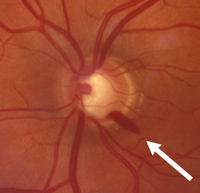
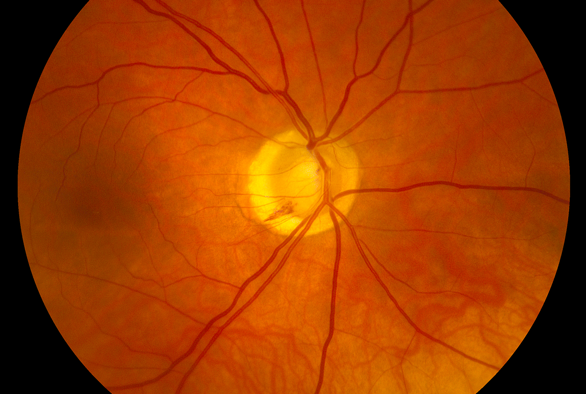
Disc photographs show enlarging cupping or new hemorrhages, while OCT shows thinning of the nerve fiber layer. A disc hemorrhage often precedes field loss, so combine structure with function.
Structure Versus Function
Structure may change before function in early disease, but in advanced disease OCT reaches a floor. Fields then become more informative, so use both across the disease course.
Intraocular Pressure and Glaucoma Progression
Higher mean pressure and large fluctuations increase risk, and the Early Manifest Glaucoma Trial showed progression falls with each millimeter of pressure reduction. Check pressure at each visit, and also reset the target when progression occurs.
Risk Factors for Fast Glaucoma Progression
Older age, thin cornea and exfoliation raise risk, and disc hemorrhages and cardiovascular disease also contribute. Pseudoexfoliation needs close care (see pseudoexfoliation syndrome), so identify these patients early.
Responding to Progression
Check adherence and technique first, then lower the target pressure with added drops, laser or surgery. Repeat fields to confirm the new course, so act on confirmed change.
Communicating With Patients
Show patients their trend lines, and explain that glaucoma is slow but can accelerate. Discuss lifestyle and treatment adherence, and also set realistic goals.
Pitfalls to Avoid
- Do not change treatment based on one field
- Never ignore reliability indices
- Avoid overlooking cataract as a cause of field loss
- Do not forget disc hemorrhages
Glaucoma Progression in Normal-Tension Disease
Patients with normal pressure can still progress, and because pressure is already low, further reduction is harder. If progression continues, look for vascular factors, so review blood pressure and sleep (see normal-tension glaucoma).
Glaucoma Progression and Cataract Surgery
Cataract surgery can lower pressure and lift field sensitivity, so when you compare fields, note the surgery date. Because the baseline changes, reset it after surgery, then continue trend analysis from the new baseline.
Glaucoma Progression and Central Fixation
Early central involvement carries a high risk to daily function, and because small central defects matter, add 10-2 testing. If central points are involved, treat more aggressively, so check the macula in all patients.
Glaucoma Progression and Life Expectancy
A slow rate may be acceptable in an older patient, but the same rate in a young patient threatens vision. Because remaining life matters, estimate the future loss and then set the target accordingly.
Glaucoma Progression and Imaging Artifacts
OCT segmentation errors can mimic change, so when images look different, review the scans manually. Because myopia affects thickness, interpret with care, and therefore confirm with the field.
Glaucoma Progression: Practical Summary
Obtain a strong baseline and repeat tests at set intervals, and if a trend appears, confirm it on repeat testing. Because adherence is a common cause, review it first, and finally adjust the target pressure.
Glaucoma Progression and Patient Education
Explain that treatment slows the disease but does not restore lost vision, and if patients understand the goal, they keep up with the drops. Because fast progressors need action, share the trend lines, then book the next test before the patient leaves.


Document what you see
Two smartphone imaging tools built for everyday clinic use — one for the slit lamp, one for the fundus.
From Choroida — the team behind this siteReferences
- Chauhan BC, Garway-Heath DF, Goni FJ, et al. Practical recommendations for measuring rates of visual field change in glaucoma. Br J Ophthalmol. 2008;92:569-573.
- Heijl A, Leske MC, Bengtsson B, et al. Reduction of intraocular pressure and glaucoma progression: results from the Early Manifest Glaucoma Trial. Arch Ophthalmol. 2002;120:1268-1279.
- Leske MC, Heijl A, Hussein M, et al. Factors for glaucoma progression and the effect of treatment: the Early Manifest Glaucoma Trial. Arch Ophthalmol. 2003;121:48-56.
- Gedde SJ, Vinod K, Wright MM, et al. Primary open-angle glaucoma preferred practice pattern. Ophthalmology. 2021;128:P71-P150.
Test yourself
A few questions straight from this article.
-
How many visual field tests are advised in the first year to build a reliable glaucoma baseline?
Two to three tests in the first year build a reliable baseline, because early fields carry more learning effect and a single test should not be judged alone. -
What question does event-based analysis of glaucoma visual field progression answer?
Event analysis flags a new defect that exceeds test variability, answering whether change happened but not how fast loss is occurring; events should be confirmed on repeat testing. -
In trend-based analysis of glaucoma progression, what does the slope of the plot represent?
Trend analysis plots the visual field index against time; the slope shows the rate of loss, and a steep negative slope means rapid progression. -
About how many visual field tests in the first two years help detect fast glaucoma progressors?
Studies suggest about six tests in the first two years help detect fast progressors, though cost and patient burden mean frequency is tailored to risk. -
Which factor can produce false glaucoma progression on visual field testing?
Cataract, small pupils, poor attention, fatigue and learning effects lower sensitivity, so reliability indices, pupil and media should always be reviewed. -
Which optic disc finding often precedes visual field loss in glaucoma?
Disc photographs show enlarging cupping or new hemorrhages, and a disc hemorrhage often precedes field loss, so structure should be combined with function. -
Why do visual fields become more informative than OCT in advanced glaucoma?
Structure may change before function in early disease, but in advanced disease OCT reaches a floor and fields then become more informative. -
What did the Early Manifest Glaucoma Trial show about intraocular pressure and progression?
The Early Manifest Glaucoma Trial showed progression falls with each millimeter of pressure reduction, so pressure is checked each visit and the target reset when progression occurs. -
When glaucoma progression is detected, what should be checked first before escalating treatment?
Adherence and technique are checked first; the target pressure is then lowered with added drops, laser or surgery, and fields repeated to confirm the new course. -
Which additional test is advised when small central defects threaten fixation in glaucoma?
Early central involvement carries a high risk to daily function, so 10-2 testing is added, and central involvement warrants more aggressive treatment.

Writes and reviews the clinical articles on Educate. Every post is checked against peer-reviewed sources before it goes live.