Corneal graft failure is the loss of clarity of a transplanted cornea.
It is the main reason for repeat keratoplasty.
Most grafts do well, but the long-term survival depends on the indication, the type of graft, and the risk profile of the host.
Knowing the pattern of failure gives the surgeon a way to look for the cause before planning another operation.
What Is Corneal Graft Failure?
Doctors consider a graft failed when it is irreversibly cloudy and does not recover with treatment.
Clinicians classify failure in time as primary failure, where the graft never clears after surgery, early failure, and late failure, which can occur years later.
Doctors also classify it by mechanism: immunologic, endothelial, infectious, mechanical, or due to recurrence of the original disease.
The distinction is important, since the management and the risk of recurrence differ.
Registry data show that survival rates are high for low-risk indications such as keratoconus and much lower for high-risk grafts (see penetrating keratoplasty).
Causes of Corneal Graft Failure
- Immune rejection: the most important cause of late failure, especially in vascularized and inflamed corneas (see corneal graft rejection)
- Endothelial failure: primary donor failure, or progressive loss of endothelial cells over time, the commonest cause of late failure in many series
- Infectious keratitis: bacterial, fungal, or herpetic, often related to sutures and steroids
- Recurrence of the original disease: herpes simplex keratitis, and some stromal dystrophies
- Glaucoma: raised pressure damages the endothelium and the graft
- Surgical trauma and wound problems: loose sutures, wound leaks, and persistent epithelial defects
- Ocular surface disease: dry eye, limbal stem cell deficiency, and exposure
All-fit Slit-Lamp Adapter
Record and share exactly what you see at the slit lamp. One adapter fits any slit lamp or surgical microscope — and any smartphone.
From Choroida — the team behind this siteRisk Factors of Corneal Graft Failure
Corneal vascularization in two or more quadrants carries one of the highest risks of rejection and failure.
A history of previous graft failure, large grafts that approach the limbus, young age of the recipient, active inflammation at the time of surgery, and herpetic eye disease all increase the risk.
Glaucoma, uveitis, and anterior synechiae are other well-known factors.
Donor factors such as the quality of the endothelium matter, but less than the recipient factors in most reports.
Evaluation of a Failing Graft
- Record the acuity, the intraocular pressure, and the presence of edema, with its distribution
- Look for keratic precipitates, a rejection line, and stromal infiltrates
- Examine the sutures for looseness and vascularization
- Evaluate the ocular surface and the tear film
- Use pachymetry and, if the view allows, specular microscopy or OCT of the anterior segment (see anterior segment OCT)
- Search for herpetic disease and infection by history and, if needed, cultures
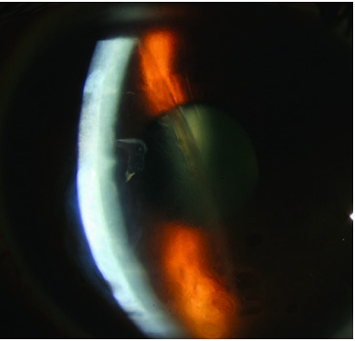
Diffuse edema with a quiet eye and a long interval since surgery suggests endothelial failure, while edema with keratic precipitates and an inflamed eye suggests rejection.
Management
Treat reversible causes first.
Rejection episodes respond to frequent topical corticosteroids, with systemic therapy for severe cases, and early treatment is essential.
Glaucoma needs control, replace loose sutures, and treat infection.
When a graft has failed permanently, options include a repeat penetrating keratoplasty, endothelial keratoplasty for isolated endothelial failure, and in selected cases a keratoprosthesis (see DSEK and DMEK and Boston keratoprosthesis).
Repeat grafts have a lower survival rate than first grafts.
The risk factors that led to the first failure often persist.
Careful preparation, control of inflammation, and treatment of the ocular surface and glaucoma before regrafting improve the odds.
Patients should understand that a second graft has a shorter expected life, and that the cause of failure influences the plan.
Preventing Failure
Close follow-up after keratoplasty identifies rejection and infection early, when treatment is most effective.
Patients should know the warning signs, which include redness, light sensitivity, and blurred vision.
They should report them immediately instead of waiting for the next appointment.
Adherence to topical steroids, and slow, supervised tapering, protects the graft, particularly in high-risk eyes.
Control of glaucoma, dry eye, and lid disease before and after surgery improves survival.
Choosing Between Procedures
Endothelial keratoplasty has lower rejection rates than full-thickness grafts and a faster visual recovery, so it is preferred for isolated endothelial failure when the stroma is clear.
When the stroma is scarred or the graft is in a vascularized bed, a repeat penetrating keratoplasty may still be required.
In eyes with multiple failures, severe surface disease, or a very high risk, a keratoprosthesis can offer visual rehabilitation, with its own long-term care requirements.
Finally, talk with the patient about the other eye and about donor tissue in advance, because the wait for a suitable graft can be long so list the patient early when a repeat graft is likely.
For the latest studies, see the PubMed results for corneal graft failure.


Document what you see
Two smartphone imaging tools built for everyday clinic use — one for the slit lamp, one for the fundus.
From Choroida — the team behind this siteReferences
- Coster DJ, Williams KA. The impact of corneal allograft rejection on the long-term outcome of corneal transplantation. Am J Ophthalmol. 2005;140:1112-1122.
- Thompson RW Jr, Price MO, Bowers PJ, Price FW Jr. Long-term graft survival after penetrating keratoplasty. Ophthalmology. 2003;110:1396-1402.
- Mannis MJ, Holland EJ, eds. Cornea. 4th ed. Elsevier; 2017.
- American Academy of Ophthalmology. Basic and Clinical Science Course, Section 8: External Disease and Cornea. AAO; current edition.
Test yourself
A few questions straight from this article.
-
When is a corneal graft considered to have failed?
Graft failure means the transplanted cornea is irreversibly cloudy and does not recover with treatment; it is the main reason for repeat keratoplasty. -
In corneal graft failure, what does the term primary failure describe?
By timing, failure is primary when the graft never clears after surgery, as distinct from early failure and late failure that can occur years later. -
Which recipient corneal finding carries one of the highest risks of graft rejection and failure?
Corneal vascularization in two or more quadrants carries one of the highest risks of rejection and failure. -
A corneal graft shows diffuse edema in a quiet eye many years after surgery. Which cause does this suggest?
Diffuse edema in a quiet eye long after surgery suggests endothelial failure, whereas edema with keratic precipitates in an inflamed eye suggests rejection. -
What is the first-line treatment of a corneal graft rejection episode?
Rejection episodes respond to frequent topical corticosteroids, with systemic therapy for severe cases, and early treatment is essential. -
A failed corneal graft has isolated endothelial failure with a clear stroma. Which procedure is preferred?
Endothelial keratoplasty has lower rejection rates and faster visual recovery than a full-thickness graft, so it is preferred for isolated endothelial failure when the stroma is clear. -
How does the survival of a repeat corneal graft compare with that of a first graft?
Repeat grafts survive less well than first grafts because the risk factors that caused the first failure often persist. -
Which option can offer visual rehabilitation in an eye with multiple corneal graft failures and severe surface disease?
In eyes with multiple failures, severe surface disease or very high risk, a keratoprosthesis can restore vision, with its own long-term care requirements. -
Which symptoms should a corneal graft recipient report immediately rather than waiting for the next visit?
Redness, light sensitivity and blurred vision are the warning signs of rejection or infection, which respond best to early treatment. -
Which indication for keratoplasty is cited as low risk, with high graft survival in registry data?
Registry data show high survival for low-risk indications such as keratoconus and much lower survival for high-risk grafts.

Writes and reviews the clinical articles on Educate. Every post is checked against peer-reviewed sources before it goes live.