AMD OCT biomarkers are features on OCT scans that help to predict progression to late disease and the response to treatment.
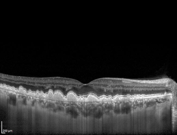
They include hyperreflective foci, drusen volume, pigment epithelial detachment, and the integrity of the outer retina.
Learning to read them improves counseling and follow-up planning.
Why AMD OCT Biomarkers Matter
Visual acuity and fundus photographs give only part of the picture.
OCT shows the retinal layers and the structures that predict future change.
Schmidt-Erfurth and Waldstein described how image-based biomarkers shape the care of neovascular disease.
AMD OCT Biomarkers in Early and Intermediate AMD
- Drusen volume and drusen regression
- Hyperreflective foci in the retina, which mark migrating RPE cells
- Subretinal drusenoid deposits, also called reticular pseudodrusen
- Loss of the ellipsoid zone and incomplete RPE and outer retinal atrophy
Nassisi and colleagues found that the number of hyperreflective foci correlates with progression within a year (see reticular pseudodrusen).
Fundus Explorer Pro
Photograph the retinal findings described here with the phone already in your pocket — 22 D optics and built-in illumination in one handheld unit.
From Choroida — the team behind this siteIncomplete Atrophy as an AMD OCT Biomarker
Christenbury and colleagues described incomplete RPE and outer retinal atrophy as a step toward geographic atrophy.
The finding warns of impending atrophy in eyes that still look intermediate on photographs.
Follow these eyes closely and counsel on the risk (see geographic atrophy treatment).
AMD OCT Biomarkers in Neovascular AMD
- Intraretinal and subretinal fluid, which guide anti-VEGF dosing
- Pigment epithelial detachment height and type
- Subretinal hyperreflective material and fibrosis
- Outer retinal tubulation and retinal atrophy
Fluid in the intraretinal space carries a different prognosis from fluid under the retina.
Using AMD OCT Biomarkers in Practice
Scan the macula in a raster pattern, and review every slice.
Record the key findings, and compare with the baseline at each visit.
Combine biomarkers with acuity and risk factors to estimate the risk of progression.
Share the findings with patients, since seeing the scan helps adherence.
Limits of AMD OCT Biomarkers
Artificial intelligence now measures some biomarkers, but validation is still under way.
Different devices give different values, so keep the same instrument.
An Example Case
A 70-year-old woman has intermediate AMD in both eyes with acuity of 20/25.
OCT in the right eye shows large drusen, several hyperreflective foci, and subretinal drusenoid deposits.
Because these features raise the risk of progression, the clinician shortens the follow-up interval to four months.
At month eight, a new neovascular membrane appears, and treatment starts early with good visual outcome.
Questions Patients Ask
Patients ask whether the spots on their scan are dangerous, and some features signal higher risk of progression.
They also ask whether anything can slow the disease, and supplements, smoking cessation, and diet may help.
Because the scans look complex, show the images and explain the key points in simple terms.
Pitfalls to Avoid
- Counting drusen on photographs only, without OCT
- Mistaking hyperreflective foci for noise in poor-quality scans
- Ignoring early outer retinal loss in an eye with good acuity
- Overtreating small amounts of fluid without a trend
Looking Ahead
Deep learning tools extract many biomarkers at once and may build risk scores that guide follow-up.
Also, trials of early interventions use these markers as outcomes.
Therefore, expect formal staging systems based on OCT.
Practical Points
Use a macular volume scan with at least 25 B-scans in each eye.
Then record the key biomarkers in the chart in a short template.
Also repeat the scan at set intervals, so that trends appear.
Anti-VEGF Response and Biomarkers
Eyes with persistent intraretinal fluid often need more frequent injections.
Large pigment epithelial detachments and thick subretinal hyperreflective material predict poorer visual outcomes.
Because patterns differ, adjust the regimen to the biomarkers.
Biomarkers and Risk Counseling
Share risk with patients in plain language.
Then give them a plan, such as home monitoring and prompt reporting of change.
Also coordinate with the primary care physician about smoking and diet.
Building a Biomarker Checklist
Write a short checklist that covers drusen size, foci, subretinal deposits, and outer retinal integrity.
Then add fluid, pigment epithelial detachment, and fibrosis for neovascular disease.
Because a template keeps reports consistent, it also helps to train residents.
Also review the checklist at every visit to find change early.
Summary
Review drusen, foci, and fluid at every visit.
Then act on change, since trends matter more than single values.
Because patients value clarity, explain the risk in plain words.
Also record the biomarkers in the same order each time.


Document what you see
Two smartphone imaging tools built for everyday clinic use — one for the slit lamp, one for the fundus.
From Choroida — the team behind this siteReferences
- Schmidt-Erfurth U, Waldstein SM. A paradigm shift in imaging biomarkers in neovascular age-related macular degeneration. Prog Retin Eye Res. 2016;50:1-24.
- Nassisi M, Fan W, Shi Y, et al. Quantity of intraretinal hyperreflective foci in patients with intermediate age-related macular degeneration correlates with 1-year progression. Invest Ophthalmol Vis Sci. 2018;59:3431-3439.
- Christenbury JG, Folgar FA, O’Connell RV, et al. Progression of intermediate age-related macular degeneration with proliferation and inner retinal migration of hyperreflective foci. Ophthalmology. 2013;120:1038-1045.
- American Academy of Ophthalmology. Basic and Clinical Science Course, Section 12: Retina and Vitreous. AAO; current edition.

Writes and reviews the clinical articles on Educate. Every post is checked against peer-reviewed sources before it goes live.