
A patient who was happy with their vision after cataract surgery returns years later complaining of fog, glare, and decreased contrast.
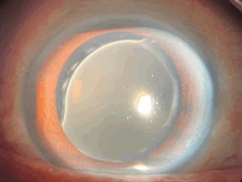
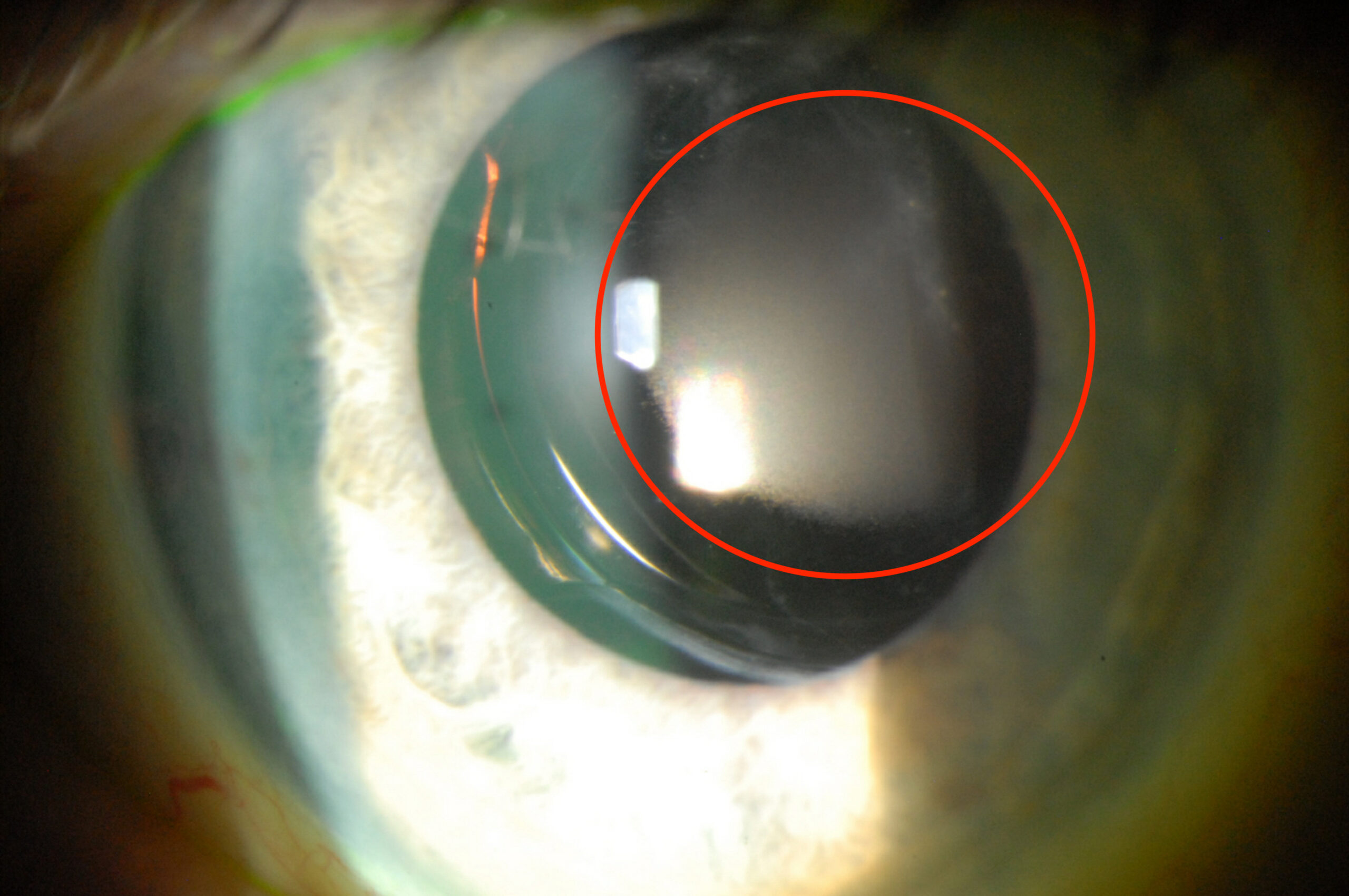
The posterior capsule looks clear, and YAG capsulotomy does not help.
If the lens implant is a hydrophilic acrylic design, the cause may be intraocular lens calcification, a rare but important complication.
What happens in the lens
Calcium phosphate crystals deposit on the surface of the lens or within its substance.
Hydrophilic acrylic lenses absorb water, and their surface chemistry makes them susceptible to precipitation of calcium and phosphate under certain conditions.
As the deposits grow, the optic becomes clouded with a granular, whitish film, and light is scattered.
Triggers and risk factors
- Specific lens materials and manufacturing batches, particularly some hydrophilic or hydrophilic-surface acrylic designs
- Breakdown of the blood-aqueous barrier, as in diabetes, uveitis, and previous glaucoma surgery
- Intraocular air or gas injection, especially in endothelial keratoplasty (Descemet stripping and DMEK), which has been linked to calcification of hydrophilic lenses in several reports
- Certain intracameral or topical drugs, such as phosphate-containing eye drops used on a damaged corneal epithelium
- Asteroid hyalosis and other conditions in which the intraocular environment is abnormal
The link with intraocular gas explains why surgeons now consider the lens type before endothelial keratoplasty in pseudophakic eyes.
All-fit Slit-Lamp Adapter
Record and share exactly what you see at the slit lamp. One adapter fits any slit lamp or surgical microscope — and any smartphone.
From Choroida — the team behind this siteSigns and symptoms
- Gradual decline in vision, months to years after surgery, in a patient with a hydrophilic lens
- Glare, haloes, and reduced contrast sensitivity
- On slit-lamp examination, a granular, white-to-gray opacity on or within the optic, which may be on the anterior surface or more diffuse
- Capsulotomy fails to improve vision because the problem is in the lens
Retroillumination shows a speckled, mottled opacity, and in some cases the deposits can be seen only after pupil dilation.
Diagnosis
Calcification is usually diagnosed from the clinical picture and the type of lens implanted.
If a surgical explant is available, histology with alizarin red or von Kossa staining, and scanning electron microscopy, confirm the presence of calcium and phosphate.
Management
- Mild changes without symptoms can be watched.
- When vision is affected, explantation and replacement of the lens is the only definitive treatment. Surgery can be hard if the capsular bag has fibrosed, and the surgeon should be prepared for a sulcus or scleral-fixated lens in some cases (see scleral-fixated intraocular lens).
- Report the case to the manufacturer and the relevant regulatory authority, with the lens batch details, to help identify safety signals.
Prevention
- Consider the lens material in patients with risk factors such as diabetes, uveitis, or planned intraocular gas
- Prefer hydrophobic acrylic IOLs in eyes likely to undergo future endothelial keratoplasty
- Record the type and model of the implanted lens in the patient’s notes and give the patient an implant card
Confirming the diagnosis
When a patient has a hydrophilic lens and a late decline in vision, the examination should show a granular, whitish opacity that follows the optic and does not change with capsulotomy. Retroillumination highlights it as a speckled pattern. Photographs help to document progression, and the operative record is needed to identify the model of the lens. If the lens is removed, send it for analysis, since staining for calcium and electron microscopy can confirm the diagnosis and help the manufacturer.
Planning the exchange
Explantation of a calcified lens can be difficult. The optic may be brittle and may need to be cut, the haptics may be fixed in the capsular bag by fibrosis, and the zonules may be weakened. Prepare for the possibility of a sulcus lens, scleral fixation, or an anterior chamber lens, and take care to preserve as much capsular support as possible. Viscoelastic, a capsular dye, and instruments that allow gentle dissection are helpful.
Informing the patient
Explain that the cloudiness is on the lens and not in the eye itself, that the opacity is unlikely to clear, and that a lens exchange gives good results in most cases. Make sure that the patient is given the name and model of the new lens, and that they are advised to inform any future surgeon about the history.
Reporting and safety
Cases of lens opacification should be reported through the local device safety system, with the model, batch number, date of implantation, and the clinical context. Collective reports allow regulators and manufacturers to identify patterns and take action. Keep any explanted lens in a suitable container for analysis, and give the patient a written record of the event.
Prognosis
After lens exchange, visual results are generally good, though explantation carries risks such as capsule damage, vitreous loss and corneal edema.


Document what you see
Two smartphone imaging tools built for everyday clinic use — one for the slit lamp, one for the fundus.
From Choroida — the team behind this siteReferences
- Neuhann IM, Kleinmann G, Apple DJ. A new classification of calcification of intraocular lenses. Ophthalmology. 2008;115:73-79.
- Werner L. Calcification of hydrophilic acrylic intraocular lenses. Am J Ophthalmol. 2008;146:341-343.
- Werner L, Apple DJ, Escobar-Gomez M, et al. Postoperative deposition of calcium on the surfaces of a hydrogel intraocular lens. Ophthalmology. 2000;107:2179-2185.
- Apple DJ, Mamalis N, Loftfield K, et al. Complications of intraocular lenses. A historical and histopathological review. Surv Ophthalmol. 1984;29:1-54.
