
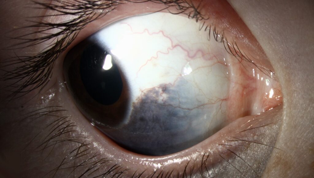
Nevus of Ota is a blue-gray patch of skin around the eye, in the distribution of the first and second divisions of the trigeminal nerve, and it is often accompanied by pigmentation of the eye itself.
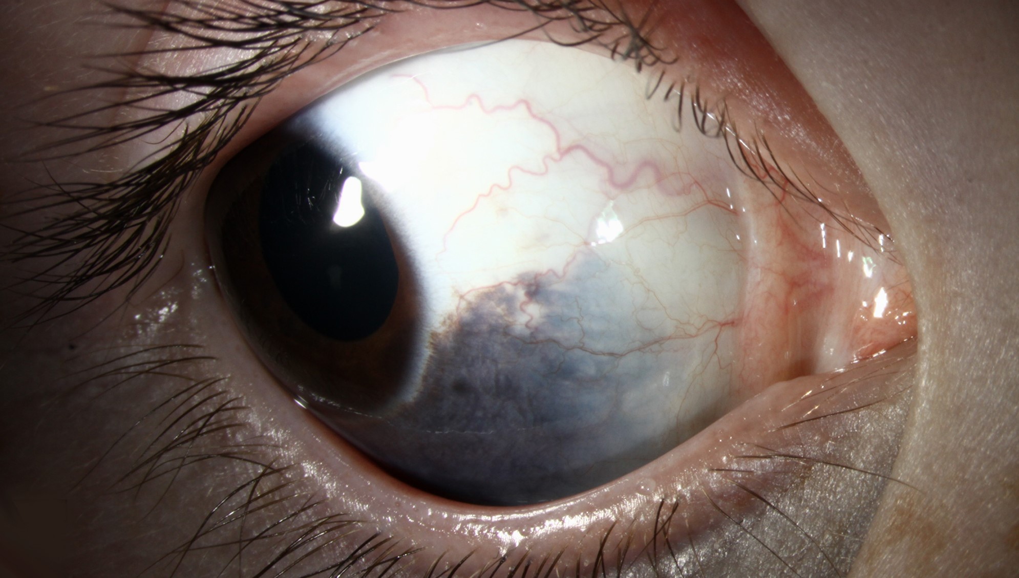
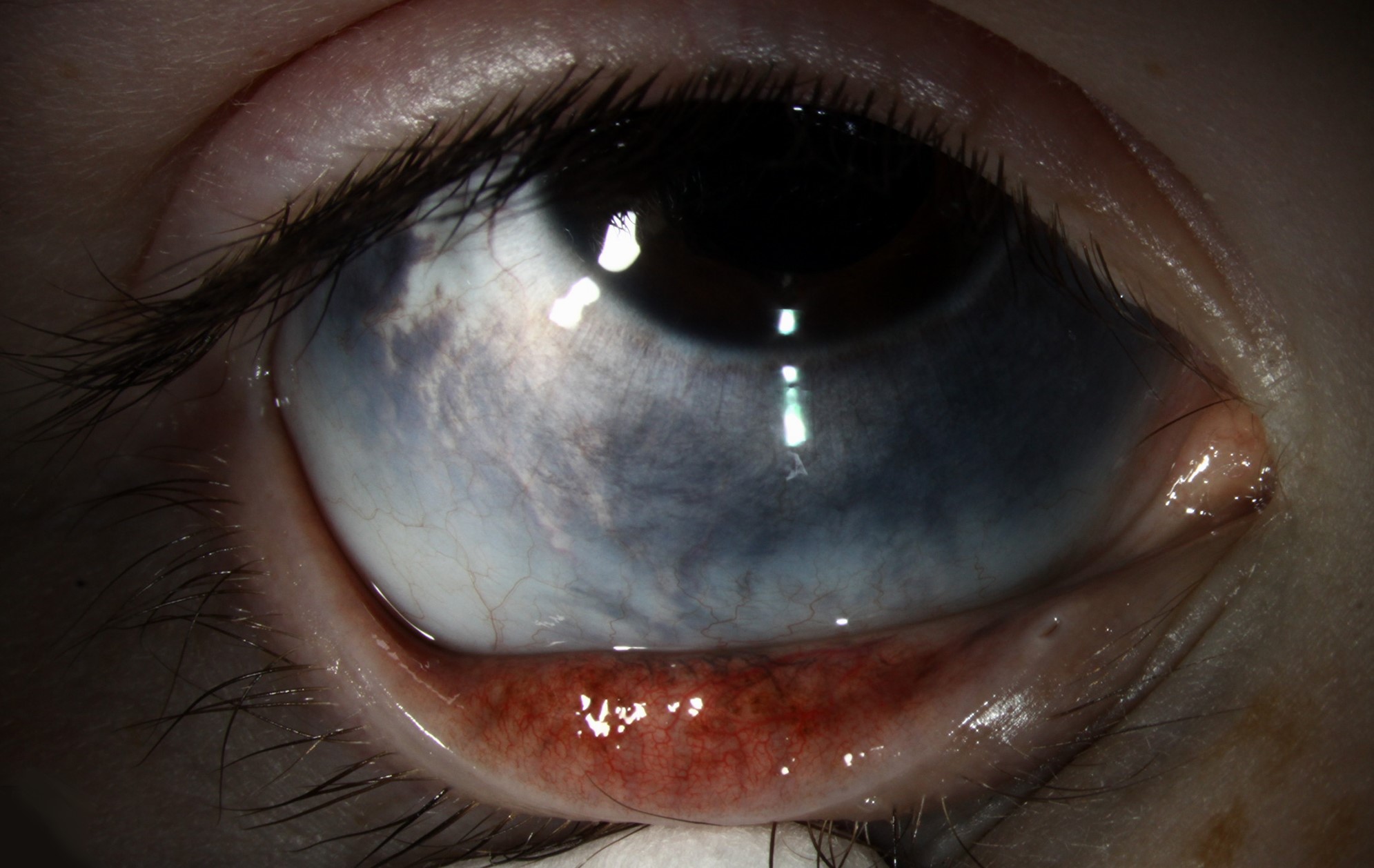
When the eye and the skin are both involved, the condition is called oculodermal melanocytosis.
The importance for the ophthalmologist is the association with uveal melanoma and glaucoma, which makes regular examination necessary.
What Is Nevus of Ota?
The nevus results from dermal melanocytes that failed to migrate completely to the epidermis during development.
The pigment is blue-gray to brown, usually unilateral, and it may be patchy or confluent, and it involves the periorbital skin, forehead, cheek, and sometimes the nose and mucous membranes.
It is most common in people of Asian and African descent, and it is more frequent in women, and in some patients it appears in childhood or at puberty.
Ocular melanocytosis refers to the increased pigmentation of the episclera, sclera, uvea, and other ocular tissues, and it may exist without a skin lesion.
Ocular Findings
- Episcleral and scleral pigment: slate-gray or blue patches that do not move with the conjunctiva
- Iris hyperpigmentation: a darker iris in the affected eye, with a mammillated surface (see iris heterochromia)
- Trabecular meshwork pigmentation: dense pigment on gonioscopy
- Choroidal thickening and a dark fundus: the fundus appears darker than in the other eye
- Optic disc pigmentation: occasionally
The pigment is not on the conjunctiva, which separates it from a conjunctival nevus or melanosis.
Fundus Explorer Pro
Photograph the retinal findings described here with the phone already in your pocket — 22 D optics and built-in illumination in one handheld unit.
From Choroida — the team behind this siteRisks
The main risks are uveal melanoma and glaucoma.
In white patients with ocular melanocytosis, the lifetime risk of uveal melanoma has been estimated at about one in 400, which is much higher than the baseline of the general population.
The risk is lower in Asian and African patients, but it is not zero (see choroidal melanoma).
Glaucoma, typically open-angle, develops in a minority of affected eyes, and it is linked to the dense pigment in the angle.
Melanoma of the orbit or brain and leptomeningeal melanocytosis are rare.
Differential Diagnosis
- Mongolian spot: a similar pigment on the sacrum or the back of infants, which usually fades
- Nevus of Ito: the same type of pigment in the supraclavicular and scapular area
- Café-au-lait macule: a lighter brown patch with a different course
- Blue nevus and melanoma: a raised, enlarging lesion
- Scleral staphyloma or thinning: an area of dark uvea showing through, with a different shape
Evaluation and Follow-Up
Examine the skin, the sclera, the iris, the angle, and the fundus under dilation.
Measure the pressure and examine the optic disc, with visual fields and OCT of the nerve fiber layer if glaucoma is suspected.
Photograph the pigment and the fundus, which provides a baseline for later changes.
Examine the patient at least once a year, because both the melanoma and the glaucoma can appear late (see iris melanoma and primary open-angle glaucoma).
Teach the patient to report a change in the vision or a change in the appearance of the eye.
Treatment
No treatment is needed for the ocular pigment.
Cosmetic treatment of the skin with Q-switched lasers is effective for many patients, and it should be done by an experienced dermatologist.
Glaucoma is treated in the usual way, and uveal melanoma needs treatment by an ocular oncology service.
How Often to Examine
A reasonable plan is an annual dilated examination with gonioscopy, measurement of the pressure, and fundus photography.
Children should be examined at least once, and a baseline photograph of the pigment and the fundus is useful for comparison.
A patient who has noticed a change in vision, a visual field defect, or a change in the pigment should be reviewed promptly.
Patients in higher risk groups, such as white patients with ocular melanocytosis, need particular care, and those with a choroidal nevus within the pigmented area should be assessed using the usual risk factors for transformation (see choroidal melanoma).
Imaging
Ultrasound or OCT can measure choroidal thickness, which is greater in the affected eye.
Wide-field photographs document the extent of the fundus pigmentation, and fundus autofluorescence can show any suspicious lesion.
In suspicious cases, B-scan ultrasound shows a solid mass, and an ocular oncology referral is made.
Counselling
Explain that the pigment is present from early life or adolescence, that it is not cancer, and that the increased risk is small but real.
Emphasize that regular examinations are the way to find a problem at an early stage.
Remind patients to protect the eyes from sun exposure and to attend every follow-up visit, because the risk continues throughout life.
Families who ask about relatives can be told that the condition is usually sporadic and not inherited in a simple pattern.


Document what you see
Two smartphone imaging tools built for everyday clinic use — one for the slit lamp, one for the fundus.
From Choroida — the team behind this siteReferences
- Teekhasaenee C, Ritch R, Rutnin U, Leelawongs N. Ocular findings in oculodermal melanocytosis. Arch Ophthalmol. 1990;108:1114-1120.
- Singh AD, De Potter P, Fijal BA, et al. Lifetime prevalence of uveal melanoma in white patients with oculo(dermal) melanocytosis. Ophthalmology. 1998;105:195-198.
- Salmon JF. Kanski’s Clinical Ophthalmology: A Systematic Approach. 9th ed. Elsevier; 2020.
- American Academy of Ophthalmology. Basic and Clinical Science Course, Section 4: Ophthalmic Pathology and Intraocular Tumors. AAO; current edition.

Writes and reviews the clinical articles on Educate. Every post is checked against peer-reviewed sources before it goes live.


