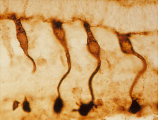
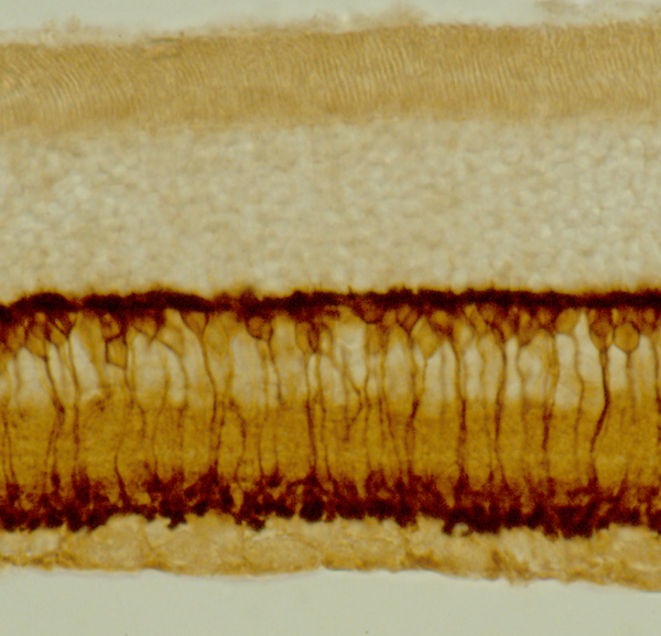
Bipolar cells carry signals from photoreceptors to ganglion cells in the retina.
They split the visual signal into ON and OFF channels for light increments and decrements.
Therefore, they form the first step of contrast coding in vision.
Where Bipolar Cells Sit
The cell bodies lie in the inner nuclear layer.
Their dendrites receive input from photoreceptors in the outer plexiform layer.
Also, their axons end in the inner plexiform layer, where they contact amacrine and ganglion cells.
As a result, they bridge the outer and inner retina.
Rod and Cone Bipolar Cells
One class of bipolar cells serves rods, and many types serve cones.
Primates have about a dozen types of cone bipolar cells.
Also, rod bipolar cells are all ON cells.
Therefore, rod and cone signals use different early circuits.
Fundus Explorer Pro
Photograph the retinal findings described here with the phone already in your pocket — 22 D optics and built-in illumination in one handheld unit.
From Choroida — the team behind this siteThe ON Pathway
ON bipolar cells depolarize when light hits the center of the receptive field.
Photoreceptors release less glutamate in light, and the cells respond through a metabotropic receptor, mGluR6.
Also, this receptor closes TRPM1 channels in the dark and opens them in light.
As a result, light increments excite ON cells.
The OFF Pathway
OFF bipolar cells hyperpolarize in light.
They use ionotropic glutamate receptors that respond directly to the transmitter.
Also, a fall in light raises glutamate release and depolarizes them.
Therefore, light decrements excite OFF cells.
Sublaminae of the Inner Plexiform Layer
OFF bipolar cells end in the outer half of the inner plexiform layer, in sublamina a.
ON bipolar cells end in the inner half, in sublamina b.
Also, this layering keeps the two channels apart down to the ganglion cells.
In addition, each ganglion cell type samples a defined depth.
Bipolar Cells and Receptive Fields
A bipolar cell has a center and a surround.
Horizontal cells provide lateral inhibition that shapes the surround.
Also, this structure enhances edges and contrast.
Therefore, retinal output signals differences and not absolute light levels.
The Rod Pathway
Rod bipolar cells do not contact ganglion cells directly.
They signal to AII amacrine cells, which connect to cone bipolar cells.
Also, AII cells use gap junctions for the ON route and glycine for the OFF route.
As a result, rod signals borrow the cone circuits in dim light.
Midget and Parasol Systems
Midget bipolar cells connect one cone to one midget ganglion cell in the fovea.
This gives high acuity and color opponency.
Also, diffuse bipolar cells feed parasol cells, which carry motion and flicker.
Therefore, the bipolar layer supports parallel processing.
The ERG b-Wave and Bipolar Cells
The b-wave of the electroretinogram reflects ON bipolar cell activity.
Müller cells convert the extracellular potassium changes into the visible wave.
Also, a normal a-wave with a reduced b-wave points to a defect after the photoreceptors.
Therefore, the electroretinogram helps to localize disease within the circuit.
Congenital Stationary Night Blindness
Complete forms involve mutations in genes such as NYX, GRM6, TRPM1, GPR179 and LRIT3.
These defects block the ON bipolar response.
Also, patients show a negative electroretinogram and poor night vision.
In addition, incomplete forms involve CACNA1F and CABP4 and affect both pathways.
Bipolar Cells in X-Linked Retinoschisis
Retinoschisin is secreted by photoreceptors and bipolar cells.
Loss of the protein disrupts cell contacts and creates cystic splits.
Also, the b-wave falls because of bipolar cell dysfunction.
Therefore, the electroretinogram is also negative in this disease.
Bipolar Cells in Retinal Degeneration
When photoreceptors die, bipolar cells lose their input.
They may rewire and form new contacts with other cells.
Also, this remodeling complicates efforts to restore vision (see retinitis pigmentosa).
As a result, optogenetic and prosthetic approaches target surviving bipolar cells.
Therapeutic Targeting
Gene therapy with light-sensitive proteins can drive ON bipolar cells.
Retinal prostheses stimulate bipolar or ganglion cells electrically.
Also, the cell type chosen changes the quality of the percept.
Therefore, circuit knowledge guides design.
Pitfalls to Avoid
- Do not equate bipolar cell dysfunction with a normal fundus and normal vision
- Never skip the electroretinogram when the history suggests night blindness
- Do not forget that rod and cone pathways differ
- Never assume that all bipolar cells depolarize in light
Horizontal and Amacrine Cells
Horizontal cells shape the surround at the first synapse.
Amacrine cells refine signals at the second synapse.
Also, they add timing and direction selectivity.
Therefore, bipolar output is only the first stage of processing.
Bipolar Cells and Vision Restoration
Optogenetic therapies express light-sensitive channels in surviving ON bipolar cells.
Electronic implants aim at the same layer.
Also, the cell type that is targeted affects contrast and motion.
As a result, outcomes differ between approaches.


Document what you see
Two smartphone imaging tools built for everyday clinic use — one for the slit lamp, one for the fundus.
From Choroida — the team behind this siteReferences
- Euler T, Haverkamp S, Schubert T, Baden T. Retinal bipolar cells: elementary building blocks of vision. Nat Rev Neurosci. 2014;15:507-519.
- Wässle H. Parallel processing in the mammalian retina. Nat Rev Neurosci. 2004;5:747-757.
- Zeitz C, Robson AG, Audo I. Congenital stationary night blindness: an analysis and update of genotype-phenotype correlations and pathogenic mechanisms. Prog Retin Eye Res. 2015;45:58-110.
- Masland RH. The neuronal organization of the retina. Neuron. 2012;76:266-280.

Writes and reviews the clinical articles on Educate. Every post is checked against peer-reviewed sources before it goes live.