Serous PED is a dome-shaped elevation of the retinal pigment epithelium (RPE) filled with clear fluid.
Clinicians often call it a fluid blister.
Although it looks simple on OCT, it can conceal neovascularization.
What a Serous PED Is
Fluid collects beneath the RPE basal lamina, above the inner collagenous layer of Bruch’s membrane.
Because the RPE lifts as one sheet, the margins look smooth and sharp.
Imaging separates it from drusenoid and vascularized types (see fibrovascular PED).
Therefore, the term describes lesion content and does not name one disease.
Why the Fluid Collects
Normally, the RPE pumps water from the retina toward the choroid.
With age, lipid accumulates in Bruch’s membrane and reduces its hydraulic conductivity.
As a result, fluid becomes trapped beneath the RPE.
In pachychoroid disease, choroidal hyperpermeability probably adds pressure from below.
Fundus Explorer Pro
Photograph the retinal findings described here with the phone already in your pocket — 22 D optics and built-in illumination in one handheld unit.
From Choroida — the team behind this siteCauses of Serous PED
Age-related macular degeneration is a common association in older patients, especially with drusen.
In younger adults, consider central serous chorioretinopathy first.
Polypoidal choroidal vasculopathy can also produce serous detachments.
In addition, Vogt-Koyanagi-Harada disease and posterior scleritis may cause them.
Symptoms Patients Report
Small extrafoveal lesions often cause no symptoms and appear during routine imaging.
When the fovea is involved, patients may notice metamorphopsia, blur or a mild hyperopic shift.
However, sudden central vision loss suggests hemorrhage, an RPE tear or active neovascularization.
Fundus Signs of Serous PED
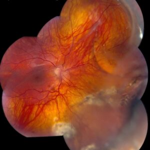
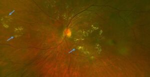
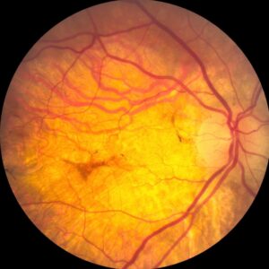
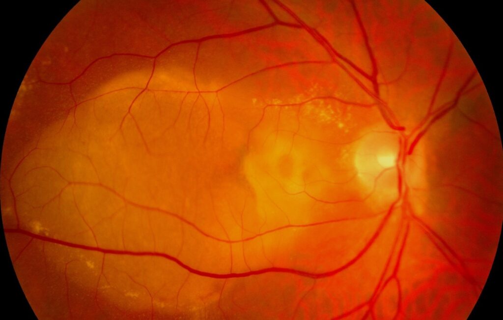
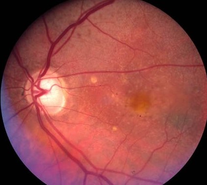
The lesion appears as a round, sharply outlined orange-yellow dome.
A faint reddish halo may surround its base.
The slit beam can also transilluminate the clear fluid, unlike blood or fibrovascular tissue.
Next, inspect the fellow eye for drusen and pigment changes.
OCT Features of Serous PED
OCT shows a smooth, steep RPE elevation over a homogeneous hyporeflective space.
Bruch’s membrane appears as a thin, flat line beneath the dome.
Because fluid transmits light well, choroidal signal often stays visible below the lesion.
By contrast, internal hyperreflective material suggests a vascular or drusenoid component.
Therefore, scan the whole lesion with dense raster volumes, since one foveal line can miss a small complex.
Fluorescein Angiography
Fluorescein fills the space early and uniformly, then pools within a stable border.
Late leakage beyond that border points away from a purely serous lesion.
Also, irregular or delayed filling should prompt a search for occult neovascularization.
ICG Angiography in Serous PED
ICG shows the dome as a hypofluorescent area.
This dark background helps reveal a hot spot, a plaque or polypoidal lesions.
Consequently, use ICG when the patient is older, the lesion looks atypical or polyps are possible.
OCT Angiography
OCT angiography offers a noninvasive look for flow beneath the RPE.
A pure fluid space should show no organized vascular signal.
However, tall domes can cause segmentation errors.
Therefore, correct the slab manually before you call a scan negative.
Excluding Hidden Neovascularization in Serous PED
First, look for a notch along the dome, a sign Gass linked to occult neovascularization.
Next, check for subretinal hemorrhage, intraretinal fluid or heterogeneous contents.
Finally, correlate any suspicious area across OCT, angiography and OCTA.
When to Observe a Serous PED
No treatment has proven benefit for an avascular lesion without active underlying disease.
Therefore, observation is reasonable when multimodal imaging shows no neovascular complex.
Some lesions flatten spontaneously.
When central serous disease is suspected, ask about corticosteroid exposure.
When to Treat
Anti-VEGF therapy is appropriate when imaging confirms macular neovascularization.
It targets the neovascular process, so it should not resolve a purely serous lesion.
Treat inflammatory causes according to the underlying disease.
Also warn patients that large vascularized detachments can develop an RPE tear.
Prognosis of Serous PED
Prognosis depends on age, cause and the presence of neovascularization.
Younger patients with central serous disease generally have a favorable course.
In elderly eyes, older series linked detachments to later neovascularization, RPE tears and atrophy.
Even after flattening, the RPE and photoreceptors may not recover fully.
Follow-Up Plan
Repeat OCT with the same device and scan pattern so that height stays comparable.
The interval depends on symptoms, fellow eye status and AMD risk.
Then repeat angiography or OCTA whenever morphology changes.
Counseling Patients
Explain that the blister is fluid under the retina’s support layer and is not a tumor.
Also explain that observation is active monitoring, because the risk of new vessels can change.
In addition, ask patients to report new distortion or sudden blur promptly.
Pitfalls to Avoid
- Do not diagnose a purely serous lesion from one OCT line
- Do not accept a negative OCTA without checking segmentation
- Avoid anti-VEGF injection for a dome without evidence of neovascularization
- Never ignore a notch, hemorrhage or heterogeneous contents


Document what you see
Two smartphone imaging tools built for everyday clinic use — one for the slit lamp, one for the fundus.
From Choroida — the team behind this siteReferences
- Mrejen S, Sarraf D, Mukkamala SK, Freund KB. Multimodal imaging of pigment epithelial detachment: a guide to evaluation. Retina. 2013;33:1735-1762.
- Zayit-Soudry S, Moroz I, Loewenstein A. Retinal pigment epithelial detachment. Surv Ophthalmol. 2007;52:227-243.
- Gass JD. Serous retinal pigment epithelial detachment with a notch: a sign of occult choroidal neovascularization. Retina. 1984;4:205-220.
- Casswell AG, Kohen D, Bird AC. Retinal pigment epithelial detachments in the elderly: classification and outcome. Br J Ophthalmol. 1985;69:397-403.
- Pauleikhoff D, Loffert D, Spital G, et al. Pigment epithelial detachment in the elderly: clinical differentiation, natural course and pathogenetic implications. Graefes Arch Clin Exp Ophthalmol. 2002;240:533-538.
- Spaide RF, Jaffe GJ, Sarraf D, et al. Consensus nomenclature for reporting neovascular age-related macular degeneration data. Ophthalmology. 2020;127:616-636.

Writes and reviews the clinical articles on Educate. Every post is checked against peer-reviewed sources before it goes live.