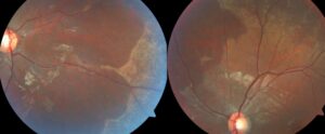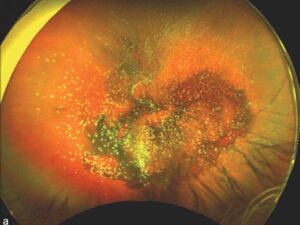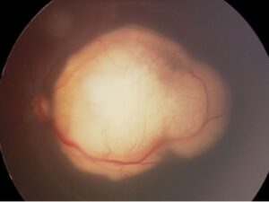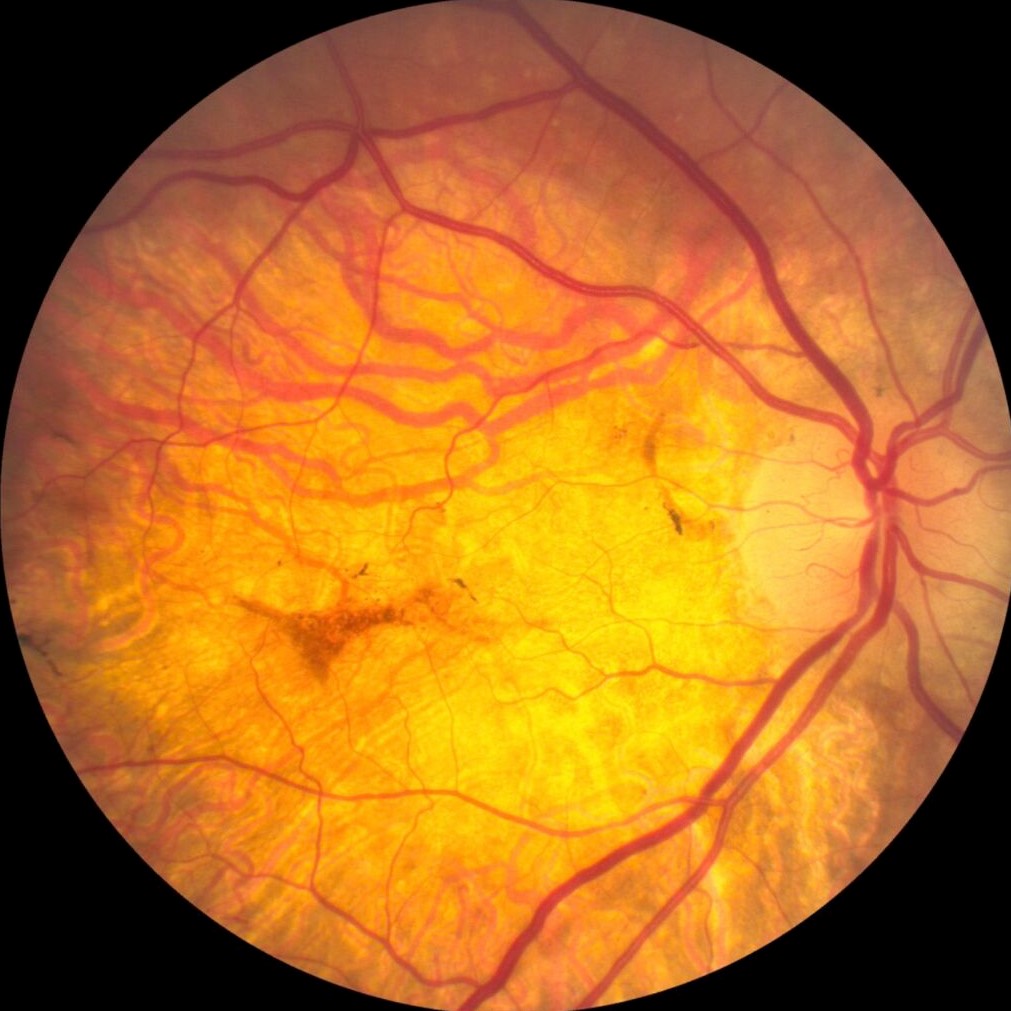
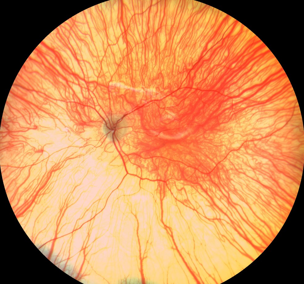
Peau d’orange fundus describes a diffuse, mottled, yellow-orange pattern at the level of the retinal pigment epithelium (RPE).
The French term means “orange skin,” because the dimpled texture resembles citrus peel.
Clinicians most often see it in pseudoxanthoma elasticum (PXE), usually alongside angioid streaks.
Therefore, this sign can prompt a systemic diagnosis before vision declines.
What Peau d’Orange Fundus Looks Like
The examiner sees fine, confluent yellowish speckling, often most obvious temporal to the macula.
Its edges usually fade gradually into the surrounding fundus.
Because the change is subtle, a quick undilated view can miss it.
Where to Look
First, inspect the temporal posterior pole and the adjacent midperiphery.
Next, compare both eyes, since PXE-related fundus changes are usually bilateral.
Fundus Explorer Pro
Photograph the retinal findings described here with the phone already in your pocket — 22 D optics and built-in illumination in one handheld unit.
From Choroida — the team behind this siteWhy Peau d’Orange Fundus Develops
PXE results from biallelic variants in the ABCC6 gene.
As a result, elastic tissues undergo progressive mineralization, including Bruch’s membrane.
Imaging work suggests that this calcification spreads outward from the posterior pole.
However, the exact histologic basis of the mottling remains incompletely defined.
Relation to Angioid Streaks
Angioid streaks are linear breaks in brittle, calcified Bruch’s membrane, and they radiate from the optic disc (see angioid streaks).
Both signs therefore reflect disease of the same layer.
Some patients show the mottling before streaks become obvious.
How Peau d’Orange Fundus Points Toward PXE
Angioid streaks also occur with Paget disease, hemoglobinopathies and other systemic disorders, and some cases remain idiopathic.
However, the combination of streaks and peau d’orange raises particular suspicion for PXE.
Still, one fundus sign cannot establish PXE by itself (see pseudoxanthoma elasticum).
Associated Fundus Signs
These findings strengthen the suspicion when they appear together:
- Angioid streaks radiating from the disc
- Comet lesions, which are small pale spots with tapering tails
- Optic disc drusen
- Choroidal neovascularization or fibrovascular scars
- Chorioretinal atrophy in advanced disease
Symptoms in Peau d’Orange Fundus
The mottling itself rarely causes symptoms.
Therefore, many patients are found during routine examination or a workup for another complaint.
Metamorphopsia or a central scotoma should suggest a macular complication.
Then, the priority shifts to excluding choroidal neovascularization.
Imaging Peau d’Orange Fundus
Near-infrared reflectance often displays the speckled texture better than color photography.
Autofluorescence adds information about RPE health and may reveal streaks or atrophy (see fundus autofluorescence).
Meanwhile, OCT shows Bruch’s membrane breaks, subretinal fluid and neovascular tissue.
Examination Pearls
First, use a dilated examination to see the mottling clearly.
Red-free light can also improve contrast for streaks and hemorrhage.
Then, document the findings with photographs so later changes are easy to compare.
Differential Diagnosis
Several conditions can resemble this pattern, so the context needs careful review:
- Fine drusen or early pattern dystrophy
- Flecked retinal disease, such as Stargardt disease
- Age-related RPE mottling
- Pigmentary retinopathies with a salt-and-pepper fundus
Systemic Workup After Peau d’Orange Fundus
First, examine the neck, axillae and flexural skin for yellowish papules.
Then, ask about claudication, gastrointestinal bleeding, early vascular disease and affected relatives.
Skin biopsy can show calcified, fragmented elastic fibers, while genetic testing can identify ABCC6 variants.
In addition, refer the patient for dermatology and cardiovascular assessment when PXE remains likely.
Monitoring the Patient
Peau d’orange itself needs no direct treatment.
However, it signals a fundus at risk, so follow-up should focus on the macula.
Home Amsler grid testing can help patients detect new distortion early.
Follow-Up Planning
First, schedule regular dilated examinations, and shorten the interval after any new visual symptom.
In addition, obtain a baseline OCT so that later fluid or new membranes are easier to recognize.
As a result, early neovascular change can be treated promptly.
Treating Neovascular Complications
Intravitreal anti-VEGF therapy is the usual treatment for choroidal neovascularization in this setting.
It suppresses leakage and can stabilize or improve vision.
However, the evidence comes mainly from case series and small prospective studies, and recurrences are common.
Treatment also carries the usual injection risks, including endophthalmitis.
It cannot reverse established atrophy, repair Bruch’s membrane or stop mineralization.
Counseling in Peau d’Orange Fundus
Explain that the sign itself rarely threatens vision.
Then explain why its possible systemic link justifies further evaluation.
Because a brittle Bruch’s membrane may tear after minor blunt trauma, advise protective eyewear and caution with contact sports.
Finally, ask patients to report new distortion promptly.
Prognosis
Long-term vision depends mainly on macular complications, including neovascularization and atrophy.
For example, a streak extending beneath the fovea carries more concern than isolated peripheral mottling.
Pitfalls to Avoid
- Do not skip the temporal fundus when examining a patient with angioid streaks
- Never diagnose PXE from fundus appearance alone
- Do not dismiss new metamorphopsia as a stable, longstanding finding
- Do not overlook the skin and vascular history


Document what you see
Two smartphone imaging tools built for everyday clinic use — one for the slit lamp, one for the fundus.
From Choroida — the team behind this siteReferences
- Finger RP, Charbel Issa P, Ladewig MS, et al. Pseudoxanthoma elasticum: genetics, clinical manifestations and therapeutic approaches. Surv Ophthalmol. 2009;54:272-285.
- Charbel Issa P, Finger RP, Götting C, et al. Centrifugal fundus abnormalities in pseudoxanthoma elasticum. Ophthalmology. 2010;117:1406-1414.
- Gliem M, Zaeytijd JD, Finger RP, et al. An update on the ocular phenotype in patients with pseudoxanthoma elasticum. Front Genet. 2013;4:14.
- Georgalas I, Papaconstantinou D, Koutsandrea C, et al. Angioid streaks, clinical course, complications, and current therapeutic management. Ther Clin Risk Manag. 2009;5:81-89.
- Clarkson JG, Altman RD. Angioid streaks. Surv Ophthalmol. 1982;26:235-246.

Writes and reviews the clinical articles on Educate. Every post is checked against peer-reviewed sources before it goes live.