Retinal microaneurysms are small, round, saccular outpouchings of retinal capillaries.
They are also the earliest clinically visible lesion of diabetic retinopathy.
Clinicians also see them in many other retinal vascular diseases, so their presence is a sign of vascular damage and not a diagnosis in itself.
Learning to distinguish them from dot hemorrhages, and to judge whether they are leaking, helps the clinician to decide when to image and when to treat.
What Are Retinal Microaneurysms?
A microaneurysm forms when a capillary wall weakens, usually after loss of pericytes, and the vessel balloons outward.
In diabetes, chronic hyperglycemia damages pericytes and endothelial cells, and the weakened wall then dilates.
Also, microaneurysms are typically 25 to 100 micrometers in diameter.
They are also most numerous in the posterior pole, temporal to the fovea.
They may also leak plasma into the surrounding retina, which forms hard exudates and macular edema, or they may thrombose and disappear.
New lesions form while old ones close, so the count changes over time, with a continuing turnover that reflects the activity of the disease.
Appearance on Examination
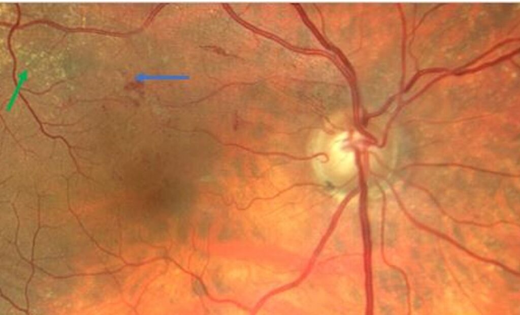
- Small, round, sharply defined red dots, often in clusters
- Intraretinal location, with no visible vessel connected to the dot
- Hard exudates arranged in a ring around a leaking cluster (a circinate pattern)
- Association with dot and blot hemorrhages and cotton wool spots in more advanced disease
It is not possible to separate a small microaneurysm from a dot hemorrhage reliably on clinical examination.
The two are grouped together in the grading of retinopathy (see diabetic retinopathy classification).
Fundus Explorer Pro
Photograph the retinal findings described here with the phone already in your pocket — 22 D optics and built-in illumination in one handheld unit.
From Choroida — the team behind this siteImaging in Retinal Microaneurysms
Fluorescein angiography shows microaneurysms as discrete hyperfluorescent dots that fill in the early phase, whereas hemorrhages block fluorescence and appear dark (see fluorescein angiography patterns).
Also, late leakage identifies those that are active, and this information guides focal laser treatment.
OCT shows microaneurysms as round hyperreflective lesions with a hyporeflective center in the inner nuclear and outer plexiform layers, with surrounding edema if they leak.
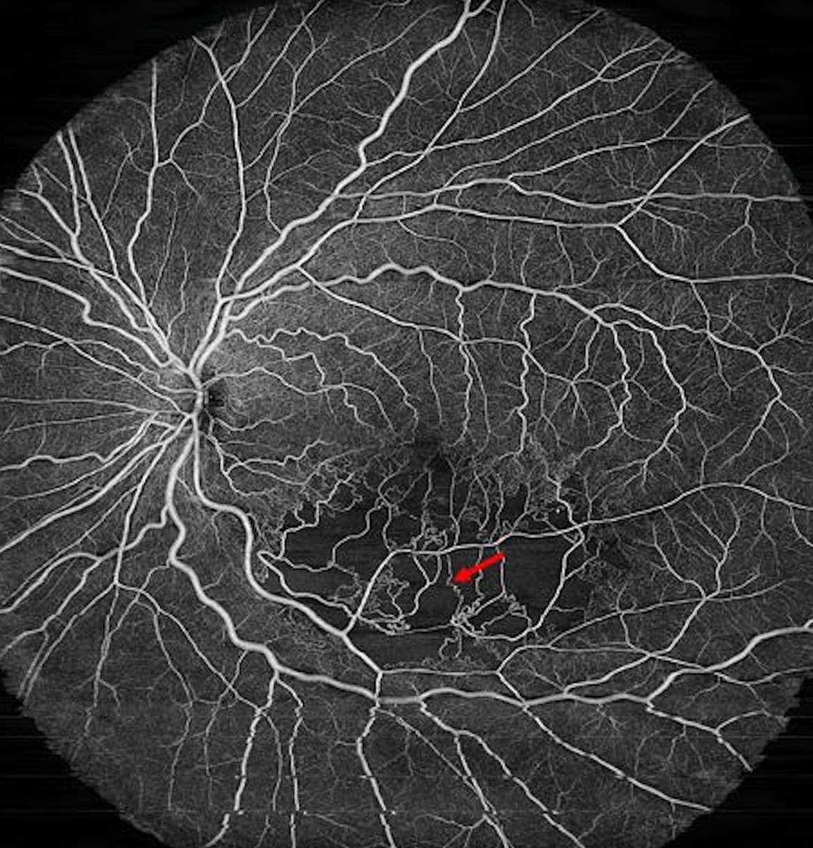
OCT angiography can display many microaneurysms without dye, although the smallest or slowly flowing ones may not be visible.
Other Conditions With Microaneurysms
- Hypertensive retinopathy and retinal vein occlusion (see central retinal vein occlusion)
- Radiation retinopathy and sickle cell retinopathy
- Macular telangiectasia and Coats disease (see Coats disease)
- Retinal arterial macroaneurysm: a larger lesion on an arteriole (see retinal macroaneurysm)
- Ocular ischemic syndrome and idiopathic juxtafoveal telangiectasis
A history of diabetes, hypertension, or a prior vein occlusion usually makes the cause clear, and unexplained microaneurysms in a young patient require a broader work-up.
Management for Retinal Microaneurysms
Microaneurysms alone do not need treatment, and the main step is systemic control of glucose and blood pressure.
Also, focal laser to leaking microaneurysms was established as a treatment for clinically significant macular edema by the ETDRS.
It reduces the risk of moderate visual loss (see diabetic macular edema).
Anti-VEGF therapy is also now preferred for center-involving macular edema, and focal laser is used for selected cases with leaking lesions at a distance from the center.
Document the count and the distribution with photographs, because an increase in the number of lesions over time signals progression.
Counselling
Tell the patient that the dots are small changes in the retinal capillaries, that they are an early sign, and that keeping glucose, blood pressure, and lipids under control reduces the chance that the disease advances.
Review the schedule for eye examinations, which should be kept even if vision is good.
Counting and Monitoring
Clinicians have used the number of microaneurysms in a standard field in research to follow the course of early retinopathy, and software can count them on photographs.
In clinical practice, the exact number matters less than the trend.
A rise in new lesions over a year predicts progression.
In addition, automated screening systems use microaneurysm detection as a key step in identifying referable disease (see AI diabetic retinopathy screening).
Pitfalls
- Confusing a drusen-like spot or a small hemorrhage with a microaneurysm
- Attributing every microaneurysm to diabetes without checking for other causes
- Treating microaneurysms on the basis of appearance alone, without OCT or angiography to show leakage
- Forgetting the fellow eye, which often shows similar changes
A mature clinician looks at the pattern as a whole, including the distribution, the associated exudates, and the findings on the macula.
When explaining the finding, a simple drawing of a capillary with a small bulge helps patients to understand what they have.
A copy of the photograph for the patient to keep reinforces the message at each visit.
For the latest studies, see the PubMed results for retinal microaneurysms.


Document what you see
Two smartphone imaging tools built for everyday clinic use — one for the slit lamp, one for the fundus.
From Choroida — the team behind this siteReferences
- Early Treatment Diabetic Retinopathy Study Research Group. Grading diabetic retinopathy from stereoscopic color fundus photographs: an extension of the modified Airlie House classification. ETDRS report number 10. Ophthalmology. 1991;98:786-806.
- Hellstedt T, Immonen I. Disappearance and formation rates of microaneurysms in early diabetic retinopathy. Br J Ophthalmol. 1996;80:135-139.
- Ryan SJ, Schachat AP, Wilkinson CP, et al, eds. Retina. 6th ed. Elsevier; 2018.
- American Academy of Ophthalmology. Basic and Clinical Science Course, Section 12: Retina and Vitreous. AAO; current edition.

Writes and reviews the clinical articles on Educate. Every post is checked against peer-reviewed sources before it goes live.