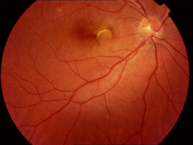
A fibrovascular pigment epithelial detachment (PED) is an elevation of the retinal pigment epithelium that contains neovascular tissue beneath it.
It is also the characteristic lesion of type 1 macular neovascularization in age-related macular degeneration.
It is also common in patients who need injections.
Its management is challenging, because the response to treatment is often incomplete and the RPE can tear.
What Is a Fibrovascular Pigment Epithelial Detachment?
The RPE sits on Bruch membrane, and in neovascular AMD, new vessels grow from the choroid into the space between them (see macular neovascularization types).
These vessels, with fibrous tissue, lift the RPE, producing the detachment.
Fluid, lipid, and blood may also accumulate in the space.
The PED may be shallow or tall, and may coexist with subretinal fluid.
Fibrovascular Pigment Epithelial Detachment: Appearance on OCT
- An irregular, undulating elevation of the RPE, often with a moderate to high internal reflectivity from the fibrovascular tissue
- A thickened or double-layer appearance of the RPE and Bruch membrane complex, called the double-layer sign
- Variable fluid above or within the PED, and possibly hyperreflective material
- Possible subretinal hyperreflective material and fibrin
- Features of the underlying disease, such as drusen
OCT angiography can reveal the flow signal of the neovascular network under the RPE (see OCT angiography).
Fundus Explorer Pro
Photograph the retinal findings described here with the phone already in your pocket — 22 D optics and built-in illumination in one handheld unit.
From Choroida — the team behind this siteDifferentiating From Other PEDs
- Serous PED: a smooth, dome-shaped elevation with homogeneous hyporeflective fluid, which occurs in central serous chorioretinopathy and in some AMD eyes (see pigment epithelial detachment)
- Drusenoid PED: a confluent soft druse, with a more homogenous content, and no neovascularization at first (see reticular pseudodrusen)
- Hemorrhagic PED: a dark, dome-shaped lesion with blood (see submacular hemorrhage)
- Polypoidal choroidal vasculopathy, with notched PEDs and polyps (see polypoidal choroidal vasculopathy)
Indocyanine green angiography and OCT angiography help to separate them.
Treatment for Fibrovascular Pigment Epithelial Detachment
- Anti-VEGF therapy: first-line, with loading doses followed by an individualized regimen (see anti-VEGF therapy)
- Treat-and-extend dosing: often used in chronic disease (see treat-and-extend regimen)
- Changing drugs: for persistent fluid or PED, changing to another agent may help (see anti-VEGF tachyphylaxis)
- Photodynamic therapy: in selected patients, such as those with polypoidal features (see photodynamic therapy)
Fibrovascular PEDs respond less than other lesions, and the height and the fluid may decrease slowly.
RPE Tear Risk
A feared complication is a tear of the RPE, which can occur spontaneously or after anti-VEGF injection, especially in large, tall PEDs.
- The risk is higher with a PED height greater than a few hundred micrometers, and with a diameter greater than about 1,000 micrometers
- A tear causes acute loss of vision if the fovea is involved, with a wave of retraction of the RPE and a bare area (see RPE tear)
- Treatment with injections should be continued if there is activity, but patients should be told of the risk
- Close monitoring after the first injections is advised
Studies suggest that anti-VEGF therapy is still beneficial in such eyes, and the risk of tear should not stop treatment.
Outcomes of Fibrovascular Pigment Epithelial Detachment
- Vision often improves less than in classic lesions, and many patients maintain stable vision
- Atrophy and fibrosis may develop over years (see geographic atrophy growth)
- Recurrent fluid and hemorrhage are common, and monitoring must continue
Counselling
Explain that the lesion is a form of wet AMD, that treatment aims to keep vision stable, and that a complete resolution of the elevation is not always possible.
Tell the patient about the small risk of RPE tear, and about the need to report any sudden loss of vision.
In addition, support the patient through the long course of treatment.
Monitoring During Treatment
Measure the height and the width of the PED on each OCT, and compare with previous scans.
A decrease in height with retention of the retinal pigment epithelium line is a good sign, while a sudden irregular collapse may signal a tear.
Look for signs of fibrosis, such as increased reflectivity, and for atrophy at the edge, since these affect the potential for recovery.
Patient Communication
Tell patients that the injections have to be given for a long time in many cases, and that the aim is to prevent loss of vision.
Make sure they know to report any sudden change in vision, such as a new dark spot, at once.
Keep records of the injection dates, the height of the PED, and the presence of fluid in the chart, so that any clinician can see the trend and adjust the interval.
Provide the patient with written information about the symptoms of RPE tear and where to seek urgent care.
For the latest studies, see the PubMed results for fibrovascular pigment epithelial detachment.


Document what you see
Two smartphone imaging tools built for everyday clinic use — one for the slit lamp, one for the fundus.
From Choroida — the team behind this siteReferences
- Spaide RF, Jaffe GJ, Sarraf D, et al. Consensus nomenclature for reporting neovascular age-related macular degeneration data: consensus on neovascular AMD nomenclature study group. Ophthalmology. 2020;127:616-636.
- Sarraf D, Chan C, Rahimy E, Abraham P. Prospective evaluation of the incidence and risk factors for the development of RPE tears after high- and low-dose ranibizumab therapy. Retina. 2013;33:1551-1557.
- Freund KB, Zweifel SA, Engelbert M. Do we need a new classification for choroidal neovascularization in age-related macular degeneration? Retina. 2010;30:1333-1349.
- American Academy of Ophthalmology. Basic and Clinical Science Course, Section 12: Retina and Vitreous. AAO; current edition.

Writes and reviews the clinical articles on Educate. Every post is checked against peer-reviewed sources before it goes live.