Dilated retinal veins are a fundus sign of raised venous pressure, thick blood, slow flow or excess flow.
Each mechanism leaves a different pattern on examination.
Therefore, distribution, laterality and associated hemorrhages guide the workup.
What Counts as Dilated Retinal Veins
The normal artery to vein caliber ratio at the disc margin is about 2:3.
Therefore, compare suspect veins with the fellow eye and with earlier photographs.
Dilation describes caliber, while tortuosity describes an abnormal course.
In contrast, venous beading means focal alternating caliber changes, typically in severe diabetic ischemia.
One vessel may show all three, so record each sign separately.
Why Dilated Retinal Veins Develop
Outflow obstruction raises pressure upstream and distends the vein.
Similarly, thick blood slows flow and increases resistance through the retinal circulation.
Reduced arterial inflow also matters, because low perfusion and hypoxia promote vasodilation.
Finally, an arteriovenous shunt delivers arterial flow directly into a vein, which enlarges it.
Fundus Explorer Pro
Photograph the retinal findings described here with the phone already in your pocket — 22 D optics and built-in illumination in one handheld unit.
From Choroida — the team behind this siteOne Eye or Both?
Unilateral dilation favors vein occlusion, carotid disease, a fistula or an arteriovenous malformation.
Bilateral dilation raises concern for hyperviscosity, severe anemia, diabetic retinopathy or papilledema.
However, carotid disease and vein occlusion can both involve two eyes, so laterality never settles the diagnosis.
Dilated Retinal Veins in Central Occlusion
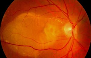
Central vein occlusion produces dilated, tortuous veins and hemorrhages in all four quadrants (see central retinal vein occlusion).
Disc edema, cotton wool spots and macular edema often accompany them.
The Central Vein Occlusion Study used 10 disc areas of nonperfusion to define ischemic disease.
Next, check for a relative afferent pupillary defect and perform undilated gonioscopy.
Branch and Hemiretinal Patterns
Branch occlusion confines dilation and hemorrhage to one sector, usually beyond an arteriovenous crossing.
Hemiretinal occlusion, by comparison, involves the superior or inferior half of the retina.
Hemorrhages fade with time, while sheathing and collateral vessels may remain.
Dilated Retinal Veins in Hyperviscosity
Hyperviscosity typically causes bilateral venous dilation with segmentation, often called sausage-linking.
Midperipheral dot and blot hemorrhages follow, and severe cases can resemble bilateral central occlusion.
Waldenström macroglobulinemia is the classic cause, although myeloma and polycythemia can also produce this picture.
Menke and colleagues linked these retinal changes to serum viscosity in Waldenström disease.
Therefore, bilateral findings with headache, mucosal bleeding or fatigue need same-day hematology contact.
Dilated Retinal Veins in Ocular Ischemia
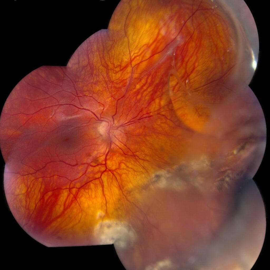
Ocular ischemia typically produces dilated veins that stay relatively straight, along with narrowed arteries.
In addition, midperipheral blot hemorrhages, aching pain and slow recovery after bright light support the diagnosis.
Also look for iris neovascularization and anterior chamber flare.
Severe ipsilateral carotid stenosis is the usual cause, so request carotid imaging promptly.
Separating Ischemia From Vein Occlusion
Hemorrhage location helps first, because ocular ischemia favors the midperiphery.
Disc edema, meanwhile, favors vein occlusion.
Angiography then adds another clue.
Ocular ischemia often delays choroidal filling and prolongs arm-to-retina time, whereas vein occlusion mainly delays venous transit.
High-Flow Dilated Retinal Veins
Retinal arteriovenous malformations connect arteries to veins without an intervening capillary bed.
Consequently, feeding and draining vessels can look similar in color and caliber.
Retinal hemangioblastoma also shows a dilated feeder artery and draining vein.
Therefore, inspect the peripheral retina and consider von Hippel-Lindau disease.
Carotid Cavernous Fistula
A fistula raises orbital venous pressure and can engorge the retinal veins.
First, examine for corkscrew episcleral vessels, proptosis, chemosis, raised intraocular pressure and a bruit.
Vascular imaging then confirms the diagnosis, because the fundus alone can mimic vein occlusion.
Workup for Dilated Retinal Veins
Match the tests to the pattern, because a fixed panel misses the clinical question.
- Blood pressure, pupils, intraocular pressure and gonioscopy
- Dilated examination with widefield photographs
- OCT for macular edema
- Fluorescein angiography for transit and nonperfusion
- Blood count, smear, glucose and lipids
- Serum viscosity and protein electrophoresis in bilateral cases
- Carotid duplex when ischemia is suspected
- Orbital and vascular imaging when a fistula is suspected
Thrombophilia panels suit only selected patients (see thrombophilia testing).
Follow-Up and Counseling
Explain that treatment targets the cause and its complications, since the dilated vein itself needs no direct therapy.
Vision then depends mainly on macular status and retinal perfusion.
Set review intervals by diagnosis and severity, and watch for macular edema and neovascularization.
Pitfalls to Avoid
- Do not label every engorged vein as central occlusion
- Never overlook hyperviscosity when findings are bilateral
- Do not skip gonioscopy in ischemic eyes
- Avoid treating dilation, tortuosity and beading as interchangeable signs


Document what you see
Two smartphone imaging tools built for everyday clinic use — one for the slit lamp, one for the fundus.
From Choroida — the team behind this siteReferences
- Central Vein Occlusion Study Group. Natural history and clinical management of central retinal vein occlusion. Arch Ophthalmol. 1997;115:486-491.
- Menke MN, Feke GT, McMeel JW, et al. Hyperviscosity-related retinopathy in Waldenström macroglobulinemia. Arch Ophthalmol. 2006;124:1601-1606.
- Mendrinos E, Machinis TG, Pournaras CJ. Ocular ischemic syndrome. Surv Ophthalmol. 2010;55:2-34.
- Brown GC, Magargal LE. The ocular ischemic syndrome: clinical, fluorescein angiographic and carotid angiographic features. Int Ophthalmol. 1988;11:239-251.
- Archer DB, Deutman A, Ernest JT, Krill AE. Arteriovenous communications of the retina. Am J Ophthalmol. 1973;75:224-241.

Writes and reviews the clinical articles on Educate. Every post is checked against peer-reviewed sources before it goes live.