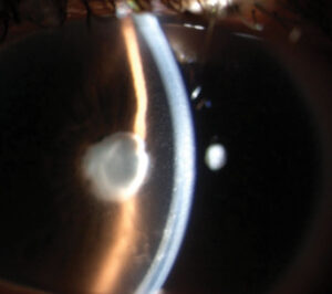
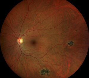
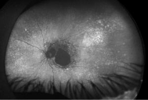
Soft drusen are larger, fuzzy deposits under the retina that raise the risk of progression to late macular degeneration.
Hard drusen are small, sharp and common with age.
Therefore, separating the two guides how closely you follow the patient.
What Drusen Are
Drusen are extracellular deposits between the retinal pigment epithelium and Bruch membrane.
They contain lipids, proteins and complement components.
Also, they are a hallmark of age-related macular degeneration (see age-related macular degeneration).
Because they are visible with a simple examination, they are an easy marker.
Hard Drusen
Hard drusen are small, round and well defined.
They measure less than about 63 micrometers.
Also, many older adults have a few of them without disease.
Therefore, isolated hard drusen carry a low risk.
Fundus Explorer Pro
Photograph the retinal findings described here with the phone already in your pocket — 22 D optics and built-in illumination in one handheld unit.
From Choroida — the team behind this siteSoft Drusen
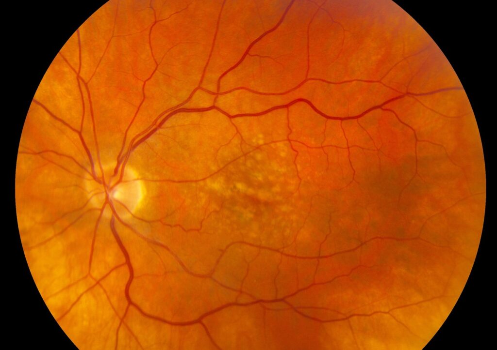
Soft drusen are larger and have indistinct, fuzzy borders.
They measure 125 micrometers or more in the common grading systems.
Also, they tend to cluster and merge.
Because they signal sub-RPE deposition, they carry a higher risk.
Intermediate Drusen
Drusen between 63 and 125 micrometers are intermediate in size.
They fall in the early stage of the Beckman classification.
Also, large drusen and pigmentary change define the intermediate stage (see Ferris classification).
Therefore, size grading decides the stage.
Why Soft Drusen Predict Disease
The Beaver Dam Eye Study found that soft indistinct drusen strongly predicted late disease.
Klein and colleagues also described the Wisconsin grading system for drusen.
Also, the risk rises with size, number and confluence.
Because the evidence is consistent, soft drusen drive the risk assessment.
Risk of Progression With Soft Drusen
In AREDS, eyes with extensive intermediate drusen or large drusen had a notable risk of advanced disease over five years.
Roughly one in five progressed in higher-risk categories.
Also, risk rises further when both eyes are affected.
Therefore, patients with soft drusen need regular follow-up.
Soft Drusen on OCT
Optical coherence tomography shows drusen as dome-shaped elevations of the RPE.
Soft drusen are larger and may have a heterogeneous core.
Also, hyperreflective foci above them signal higher risk (see fundus autofluorescence).
Because OCT adds detail, use it to stage and follow disease.
Other Drusen Types
Reticular pseudodrusen sit above the RPE and carry a distinct risk (see reticular pseudodrusen).
Pachydrusen occur in thick choroid eyes (see pachydrusen).
Also, basal laminar drusen look different on angiography (see basal laminar drusen).
Therefore, name the type on the report.
Differential Diagnosis
Hereditary drusen appear early and in a radial pattern (see autosomal dominant drusen).
Peripheral drusen are common and have a low risk (see peripheral drusen).
Also, inflammatory lesions may mimic drusen.
Because history matters, consider the age and the family.
Management of Soft Drusen
Advise stopping smoking and eating a healthy diet.
Offer supplements according to AREDS2 for intermediate disease.
Also, teach home monitoring with an Amsler grid and review any change at once.
Because early treatment of wet disease preserves vision, educate the patient.
Follow-Up Interval for Soft Drusen
Review patients with soft drusen every six to twelve months.
Shorten the interval if the fellow eye has late disease.
Also, repeat OCT at each visit.
Therefore, a plan helps catch change early.
Counseling
Explain that drusen are common and that most patients keep good vision.
Also, explain the signs that need a prompt visit.
Because fear is common, give reassurance with a clear plan.
Soft Drusen and Pigmentary Change
Pigment clumping over drusen increases the risk further.
Hypopigmentation also marks the intermediate stage.
Also, the combination of large drusen and pigment change defines high risk.
Because the combination matters, grade both.
Genetic and Environmental Factors
Variants in complement factor H and ARMS2 influence the risk of progression.
Smoking and diet also matter.
Also, family history increases the risk.
Therefore, ask about all three in your history.
Drusen Regression
Drusen may shrink or disappear with time.
Collapse can leave atrophy behind.
Also, regression is not always a good sign.
Because the process is complex, follow the OCT.
Visual Symptoms
Many patients with soft drusen have good acuity.
Some report slow dark adaptation or reduced contrast.
Also, metamorphopsia suggests neovascular change.
Therefore, check an Amsler grid at each visit.
Communication With the Patient
Show the drusen on the fundus photograph.
Explain the difference between small and large deposits.
Also, give written advice on lifestyle and symptoms.
For the latest studies, see the PubMed results for soft drusen.


Document what you see
Two smartphone imaging tools built for everyday clinic use — one for the slit lamp, one for the fundus.
From Choroida — the team behind this siteReferences
- Bird AC, Bressler NM, Bressler SB, et al. An international classification and grading system for age-related maculopathy and age-related macular degeneration. Surv Ophthalmol. 1995;39:367-374.
- Klein R, Davis MD, Magli YL, Segal P, Klein BE, Hubbard L. The Wisconsin age-related maculopathy grading system. Ophthalmology. 1991;98:1128-1134.
- Ferris FL 3rd, Wilkinson CP, Bird A, et al. Clinical classification of age-related macular degeneration. Ophthalmology. 2013;120:844-851.
- Klein R, Klein BE, Knudtson MD, Meuer SM, Swift M, Gangnon RE. Fifteen-year cumulative incidence of age-related macular degeneration: the Beaver Dam Eye Study. Ophthalmology. 2007;114:253-262.

Writes and reviews the clinical articles on Educate. Every post is checked against peer-reviewed sources before it goes live.