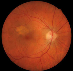
CSME, or clinically significant macular edema, is a slit lamp diagnosis defined by the Early Treatment Diabetic Retinopathy Study (ETDRS).
It describes retinal thickening or hard exudates near the macular center.
Although OCT now guides practice, the term still appears in referrals and examinations.
Therefore, clinicians should know how it translates into current language.
What CSME Means
ETDRS investigators used this definition to identify edema that threatened or involved the fovea.
It describes location, not symptoms.
As a result, a patient with 20/20 vision can still have CSME.
The Three ETDRS Criteria for CSME
An eye qualifies when it shows any one of these findings:
- Retinal thickening at or within 500 microns of the macular center
- Hard exudates at or within 500 microns of the center, with adjacent retinal thickening
- A zone of thickening of at least 1 disc area, with any part within 1 disc diameter of the center
Fundus Explorer Pro
Photograph the retinal findings described here with the phone already in your pocket — 22 D optics and built-in illumination in one handheld unit.
From Choroida — the team behind this siteThickening and Exudates
The first criterion places edema close to the fovea, though it need not reach the exact center.
For example, edema 400 microns away qualifies.
For the second criterion, exudates alone are not enough, because the adjacent retina must also be thickened.
As a result, residual exudates left after the edema resolves do not qualify.
The One Disc Area Rule
The third criterion captures larger zones farther from the center.
One disc diameter measures about 1500 microns, so the optic disc works as a ruler.
Therefore, an eye can qualify even when the fovea looks uninvolved.
Examining for CSME at the Slit Lamp
Dilate the pupil, then use a stereoscopic view with a 78 D, 90 D or contact lens (see slit lamp examination technique).
Direct ophthalmoscopy lacks stereopsis, so it cannot reliably detect thickening.
Finally, record the location of the edema relative to the center.
ETDRS Laser Results in CSME
The ETDRS compared focal photocoagulation with deferred treatment in eyes with mild to moderate nonproliferative retinopathy.
At 3 years, laser reduced moderate visual loss by about half.
However, few eyes gained substantial acuity, and laser can cause paracentral scotoma (see focal and grid laser).
From CSME to OCT Terms
Current trials classify edema by center involvement on OCT.
The ETDRS grid has a central subfield 1 mm in diameter, so its radius matches the 500 micron distance in the first criterion.
- First criterion at the center: usually center-involved edema
- First criterion off center: possibly non-center-involved edema, because the subfield average may stay normal
- Second criterion: hyperreflective deposits with fluid in the adjacent retina
- Third criterion: a thickened zone, often outside the central subfield
OCT Thickness Thresholds
Normal values vary by device and sex, so no universal cutoff exists.
For example, DRCR Protocol V used 320 microns for men and 305 for women on Spectralis.
Therefore, read thickness against the correct device reference.
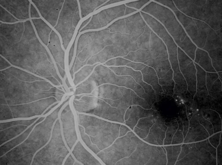
When CSME and OCT Disagree
OCT may show thickening that biomicroscopy misses.
Conversely, a normal central subfield value can hide a focal off-center lesion.
Because of this, review the full thickness map and individual scans.
Treatment Decisions Today
Modern treatment depends on center involvement and visual acuity.
Protocol T favored aflibercept at 1 year when baseline acuity was 20/50 or worse.
Meanwhile, Protocol V found no significant 2 year difference in vision loss among aflibercept, laser and observation with 20/25 or better acuity (see DME treatment choices).
Excluding Other Causes
Epiretinal membrane, vitreomacular traction, vein occlusion and cystoid edema can coexist with retinopathy.
OCT often reveals these causes.
As a result, the expected response to treatment may change.
Systemic Factors That Worsen Edema
Fluid retention from renal disease, pregnancy or glitazone use may aggravate macular edema.
Therefore, ask about medications and kidney function at the first visit.
Also, poor blood pressure control can sustain vascular leakage.
As a result, coordinate care with the patient’s physician.
Follow-Up After Treatment
Schedule visits according to the treatment chosen, because injection and laser protocols differ.
Then compare OCT thickness and acuity with the previous visit.
In addition, look for new hemorrhages or neovascularization, since retinopathy can progress during treatment.
Counseling Patients With CSME
Explain that the term describes where the swelling lies, so good vision does not exclude it.
Next, discuss that injections often require repeated visits.
Also, review glucose, blood pressure and lipid control.
Finally, advise prompt review for new blur or distortion.
Pitfalls to Avoid
- Do not treat CSME and center-involved edema as identical terms
- Do not count residual exudates without adjacent thickening
- Avoid relying on central subfield thickness alone
- Never use direct ophthalmoscopy to exclude thickening
- Do not treat on the label alone, because acuity guides the plan


Document what you see
Two smartphone imaging tools built for everyday clinic use — one for the slit lamp, one for the fundus.
From Choroida — the team behind this siteReferences
- Early Treatment Diabetic Retinopathy Study Research Group. Photocoagulation for diabetic macular edema. ETDRS report number 1. Arch Ophthalmol. 1985;103:1796-1806.
- Browning DJ, McOwen MD, Bowen RM, O’Marah TL. Comparison of the clinical diagnosis of diabetic macular edema with diagnosis by optical coherence tomography. Ophthalmology. 2004;111:712-715.
- Wells JA, Glassman AR, Ayala AR, et al. Aflibercept, bevacizumab, or ranibizumab for diabetic macular edema. N Engl J Med. 2015;372:1193-1203.
- Baker CW, Glassman AR, Beaulieu WT, et al. Aflibercept vs laser vs observation for center-involved diabetic macular edema with good visual acuity. JAMA. 2019;321:1880-1894.

Writes and reviews the clinical articles on Educate. Every post is checked against peer-reviewed sources before it goes live.