Paton lines are curved retinal folds around a swollen optic disc, classically seen in papilledema.
Leslie Paton and Gordon Holmes described them in their 1911 histological study of papilledema, and today OCT shows them far more clearly than the ophthalmoscope. Recognizing them helps confirm true disc edema and judge its severity.
What Paton Lines Look Like
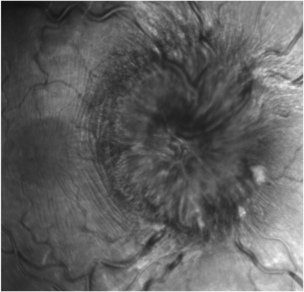
On fundus examination, Paton lines appear as fine, curved, parallel striations next to the disc margin, where they run circumferentially and follow the contour of the swollen nerve head. The temporal side usually shows them best, and red-free light makes them easier to see.
Why the Folds Form
Axoplasmic stasis swells the nerve fiber layer at the disc, and as a result the expanding nerve head pushes the adjacent retina outward. The peripapillary retina has little room to move, so it buckles into folds. Raised pressure in the nerve sheath may also deform the peripapillary Bruch membrane and add mechanical stress.
Fundus Explorer Pro
Photograph the retinal findings described here with the phone already in your pocket — 22 D optics and built-in illumination in one handheld unit.
From Choroida — the team behind this sitePaton Lines on OCT
OCT B-scans through the disc margin show the folds as undulations of the retinal layers. In many eyes, the wrinkling involves the inner retina and the outer nuclear layer. Meanwhile, the retinal pigment epithelium under these folds usually stays smooth, and therefore OCT helps separate them from folds that start in the choroid.
The IIHTT OCT Substudy
Sibony and colleagues analyzed folds in patients from the Idiopathic Intracranial Hypertension Treatment Trial and described peripapillary wrinkles, retinal folds and choroidal folds as separate patterns. OCT detected folds that fundus photographs often missed, and folds also tended to accompany more severe disc swelling.
Paton Lines vs Choroidal Folds
Choroidal folds involve the choroid, Bruch membrane and RPE, and clinically they appear as broader alternating light and dark bands. They often cross the posterior pole horizontally or obliquely (see choroidal folds), while Paton lines, by contrast, stay close to the disc and follow its edge.
Imaging Clues That Separate Them
Fluorescein angiography shows choroidal folds as alternating hyperfluorescent and hypofluorescent stripes, while Paton lines lack this pattern because the RPE remains flat. On OCT, choroidal folds show a wavy RPE and Bruch line over an undulating choroid, and enhanced depth imaging also makes the choroidal contour easier to judge.
Can Both Appear Together?
Both fold types can coexist in papilledema, and in this setting choroidal folds may reflect pressure transmitted to the posterior globe. However, choroidal folds also occur in hyperopia, orbital masses, scleritis and hypotony, so their presence alone does not prove raised intracranial pressure.
What Paton Lines Tell You About Disc Swelling
These folds point to genuine edema of the nerve head, so they help separate swollen discs from pseudopapilledema, where such folds are not expected. Buried drusen and crowded discs usually lack them (see optic disc drusen), but their absence does not exclude papilledema.
Paton Lines in Other Disc Edema
These folds are not specific to raised intracranial pressure, and they may appear with any marked disc swelling, including ischemic optic neuropathy and optic neuritis. Therefore, they confirm swelling, while the cause still needs its own workup, and a structured approach to disc edema helps (see optic disc swelling causes).
Symptoms and Paton Lines
Most patients have no symptoms from the folds themselves, and vision in papilledema usually depends on optic nerve function. However, folds that extend toward the fovea may cause metamorphopsia or blur, and then macular OCT clarifies whether folds or fluid explain the complaint.
Examining for Paton Lines
First, examine the disc stereoscopically at the slit lamp with a high plus lens, and next use red-free light and inspect the temporal margin. Then, obtain OCT line scans perpendicular to the disc edge, and finally take fundus photographs for later comparison.
Paton Lines at Follow-Up
Folds tend to fade as disc swelling resolves, and as a result serial OCT can track the response to treatment of the underlying cause. Some folds may persist for a while after the disc settles, so interpret a residual fold alongside RNFL thickness and visual function.
Talking to the Patient
Explain that the lines are wrinkles in the retina caused by nerve swelling and rarely harm vision by themselves. Stress that the cause of the swelling matters most, so timely workup and repeat imaging stay essential.
Pitfalls to Avoid
- Calling every peripapillary fold a choroidal fold
- Absent folds do not prove pseudopapilledema
- Folds do not always mean raised intracranial pressure
- Skipping OCT when the fundus view is unclear
- Forgetting a macular scan when vision drops


Document what you see
Two smartphone imaging tools built for everyday clinic use — one for the slit lamp, one for the fundus.
From Choroida — the team behind this siteReferences
- Paton L, Holmes G. The pathology of papilloedema: a histological study of sixty eyes. Brain. 1911;33:389-432.
- Sibony PA, Kupersmith MJ, Feldon SE, et al. Retinal and choroidal folds in papilledema. Invest Ophthalmol Vis Sci. 2015;56:5670-5680.
- Sibony P, Kupersmith MJ, Rohlf FJ. Shape analysis of the peripapillary RPE layer in papilledema and ischemic optic neuropathy. Invest Ophthalmol Vis Sci. 2011;52:7987-7995.
- Friedman DI, Liu GT, Digre KB. Revised diagnostic criteria for the pseudotumor cerebri syndrome in adults and children. Neurology. 2013;81:1159-1165.
- American Academy of Ophthalmology. Basic and Clinical Science Course, Section 5: Neuro-Ophthalmology. San Francisco: AAO; 2023.
Test yourself
A few questions straight from this article.
-
What are Paton lines?
Paton lines are curved folds of the retina lying around a swollen optic disc, and they are classically seen in papilledema. -
What mechanism produces Paton lines around a swollen optic disc?
Axoplasmic stasis swells the nerve fiber layer at the disc, and the enlarging nerve head pushes the peripapillary retina outward. With little room to move, that retina buckles into folds. -
Which fundus examination approach best brings out Paton lines?
The temporal side of the disc usually shows Paton lines best, and red-free light makes these fine circumferential striations easier to see. -
On OCT B-scans through Paton lines, which layer usually stays smooth beneath the folds?
The wrinkling of Paton lines often involves the inner retina and outer nuclear layer while the RPE usually stays smooth, which helps separate them from folds that start in the choroid. -
Which fluorescein angiography pattern identifies choroidal folds rather than Paton lines?
Choroidal folds show alternating hyperfluorescent and hypofluorescent stripes on angiography. Paton lines lack this pattern because the RPE stays flat. -
What do Paton lines around an elevated optic disc favor?
These folds point to true edema of the nerve head. They are not expected in pseudopapilledema, and buried drusen and crowded discs usually lack them. -
How should the absence of Paton lines be read when papilledema is suspected?
Folds support true swelling when present, but their absence neither excludes papilledema nor proves pseudopapilledema. -
Besides papilledema, in which conditions may Paton lines appear with marked disc swelling?
Paton lines are not specific to raised intracranial pressure and may appear with any marked disc swelling. They confirm swelling, while the cause still needs its own workup. -
In the Idiopathic Intracranial Hypertension Treatment Trial OCT analysis, what did folds tend to accompany?
Sibony and colleagues found that folds tended to accompany more severe disc swelling, and OCT detected folds that fundus photographs often missed. -
How should a residual Paton line be interpreted after optic disc swelling has settled?
Folds tend to fade as swelling resolves, but some persist for a while after the disc settles. A residual fold is therefore read together with RNFL thickness and visual function.

Writes and reviews the clinical articles on Educate. Every post is checked against peer-reviewed sources before it goes live.