
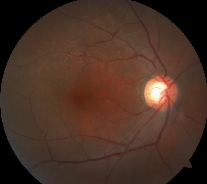
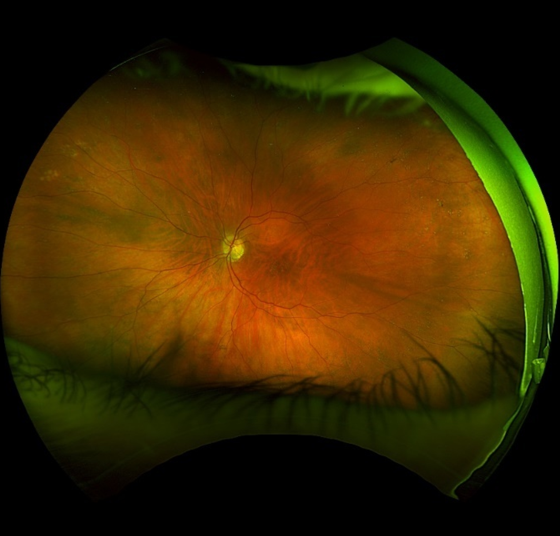
Peripheral retinal drusen look, under direct observation, essentially identical to the drusen found in the macula, small, round, yellow-white deposits scattered beneath the retina, but their location alone completely changes their clinical meaning, since drusen confined to the periphery carry none of the disease-risk implications that the very same-looking deposits carry when found in the macula.
What Drusen Actually Are
Drusen represent extracellular deposits accumulating between the retinal pigment epithelium and Bruch membrane, and while their composition and appearance can be similar regardless of location, it is specifically macular drusen that serve as an important early marker for age-related macular degeneration and its associated risk of future central vision loss.
Why Location Changes Everything
Peripheral retinal drusen are a common, essentially universal age-related finding, increasing in prevalence with advancing age, and their presence in the peripheral retina alone, without macular involvement, is not associated with an increased risk of age-related macular degeneration or any other significant visual threat, representing instead a benign, expected accompaniment of normal aging in the peripheral fundus.
Macular drusen, in sharp contrast, particularly larger, soft, confluent drusen, are a recognized risk factor and early sign of age-related macular degeneration, a leading cause of central vision loss in older adults, and their presence prompts a fundamentally different level of attention, monitoring, and patient counseling.
Fundus Explorer Pro
Photograph the retinal findings described here with the phone already in your pocket — 22 D optics and built-in illumination in one handheld unit.
From Choroida — the team behind this siteDistinguishing Features
- Location is the single most important distinguishing factor: drusen confined to the peripheral retina, well outside the macular region, carry the benign implication, while any drusen within the macula itself carry the more significant implication regardless of their individual appearance
- Peripheral drusen tend to be smaller and more uniform (hard drusen) in general, though this is a less reliable distinguishing feature than location alone, since macular drusen can also be hard and small, particularly in earlier stages
- Macular soft drusen, larger with indistinct, poorly demarcated borders, and drusen confluence (adjacent drusen merging together) are additional macular-specific features carrying particular significance for age-related macular degeneration risk, findings without the same implication when they occur, less commonly, outside the macula
Clinical Examination
- A complete dilated fundus examination should always specifically assess and document the macula separately from the peripheral retina, since the same descriptive term, drusen, carries such different weight depending on exactly where in the fundus it is used
- Careful examination of the macula for drusen size, number, confluence, and any associated pigmentary change is central to age-related macular degeneration risk assessment and staging
- Peripheral drusen, once recognized as such and confirmed to be an isolated peripheral finding without macular involvement, require no specific documentation beyond routine, standard fundus examination notes
Clinical Significance for Patients
- A patient found to have peripheral drusen alone, without macular findings, can be reassured that this specific finding does not indicate any elevated risk for age-related macular degeneration or need any specific monitoring beyond routine, age-appropriate eye care
- A patient found to have macular drusen, by contrast, benefits from appropriate counseling about age-related macular degeneration risk, monitoring recommendations including home Amsler grid monitoring for symptom changes, discussion of modifiable risk factors such as smoking cessation, and, depending on drusen characteristics and any associated findings, consideration of specific nutritional supplementation shown in clinical trials to reduce progression risk in appropriately selected patients
Avoiding Confusion in Clinical Communication
Because the term “drusen” alone, without specifying location, can be ambiguous and potentially alarming if a patient searches the term without understanding this crucial distinction, clear communication specifying “peripheral” versus “macular” drusen, and what each specifically means for that patient’s individual risk, supports both accurate understanding and appropriate reassurance or vigilance as the specific finding warrants.
Prognosis
Peripheral retinal drusen, confirmed to be isolated from the macula, carry an excellent prognosis and no specific disease implications, remaining simply a benign, common accompaniment of ocular aging.
Macular drusen, in contrast, warrant ongoing monitoring and risk-factor-based counseling given their established association with age-related macular degeneration, underscoring why the seemingly simple act of noting exactly where drusen are located during a dilated fundus examination carries such real clinical weight.


Document what you see
Two smartphone imaging tools built for everyday clinic use — one for the slit lamp, one for the fundus.
From Choroida — the team behind this siteReferences
- Age-Related Eye Disease Study Research Group. A randomized, placebo-controlled, clinical trial of high-dose supplementation with vitamins C and E, beta carotene, and zinc for age-related macular degeneration and vision loss. Arch Ophthalmol. 2001;119:1417-1436.
- Bird AC, Bressler NM, Bressler SB, et al. An international classification and grading system for age-related maculopathy and age-related macular degeneration. Surv Ophthalmol. 1995;39:367-374.
- Klein R, Klein BE, Knudtson MD, Meuer SM, Swift M, Gangnon RE. Fifteen-year cumulative incidence of age-related macular degeneration. Ophthalmology. 2007;114:253-262.
- Age-Related Eye Disease Study 2 Research Group. Lutein + zeaxanthin and omega-3 fatty acids for age-related macular degeneration. JAMA. 2013;309:2005-2015.
