Retinal vasculitis treatment depends on one early decision: is the inflammation infectious or immune mediated?
Steroids given to an eye with syphilis, tuberculosis or viral retinitis can cause rapid harm. Therefore, every plan begins with targeted testing and then escalates in steps.
Goals of Retinal Vasculitis Treatment
Treatment aims to stop active leakage, prevent vessel occlusion and protect the macula, and it also aims to limit cumulative steroid exposure over years of disease. However, no drug restores retina that is already ischemic, so early control usually achieves more than later rescue.
Step 1: Exclude Infection First
First, confirm the vascular pattern and the likely cause (see the diagnostic approach), and note that baseline tests usually include syphilis serology, an interferon gamma release assay and chest imaging. Next, consider HIV testing, toxoplasma serology and aqueous PCR when retinitis is present. Then treat any infection with specific antimicrobials before adding immunosuppression.
Fundus Explorer Pro
Photograph the retinal findings described here with the phone already in your pocket — 22 D optics and built-in illumination in one handheld unit.
From Choroida — the team behind this siteAssessing Severity Before Starting
Severity guides the intensity of therapy, so mild peripheral sheathing with good acuity may only need observation. In contrast, macular edema, occlusive disease or bilateral involvement needs prompt systemic treatment.
Step 2: Systemic Corticosteroids
Oral prednisone is the usual first systemic agent for noninfectious disease, and many specialists start near 1 mg/kg/day, while severe cases may need intravenous methylprednisolone. The dose then tapers slowly once leakage settles on angiography, because rapid tapering often triggers a rebound flare.
Steroid Taper in Retinal Vasculitis Treatment
A taper usually spans weeks to months and follows activity, not the calendar, so repeat angiography before each major dose reduction. Expert guidelines suggest reaching a low daily maintenance dose within roughly three months, and if that target fails, add a steroid-sparing agent.
Steroid Side Effects to Discuss
Patients should hear about the risks before the first tablet, and common problems include weight gain, mood change, raised glucose and insomnia. Over time, osteoporosis, cataract and ocular hypertension also appear, so arrange bone protection and regular blood pressure and glucose checks.
Step 3: Steroid-Sparing Agents
Antimetabolites such as methotrexate, mycophenolate and azathioprine form the next tier (see steroid-sparing immunosuppression). Calcineurin inhibitors such as cyclosporine are also an option, especially in Behçet disease. These drugs need several weeks to months to reach full effect, so start them early in sight-threatening or chronic disease.
Step 4: Biologics in Retinal Vasculitis Treatment
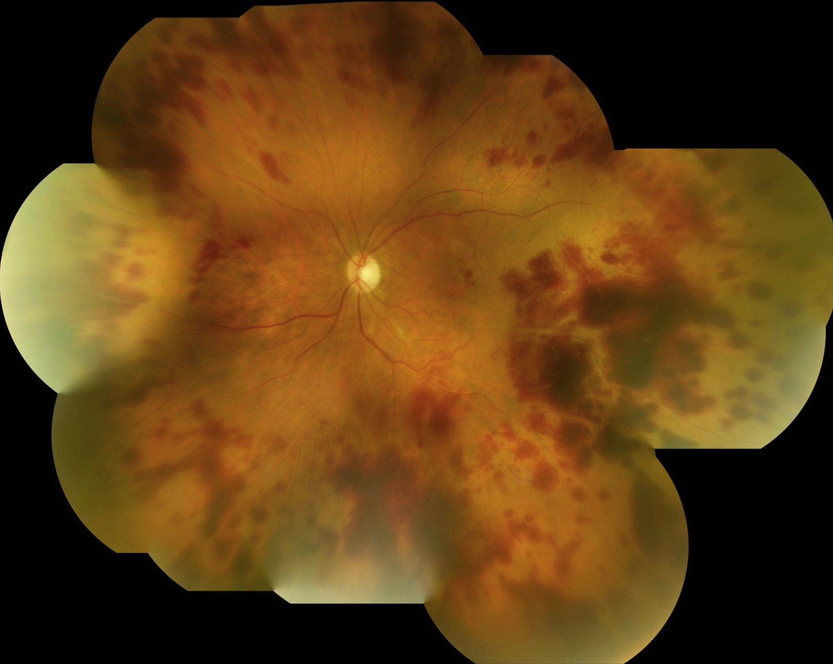
Anti-TNF agents are the main biologic option, and in the VISUAL trials, adalimumab reduced treatment failure in active and inactive noninfectious uveitis. Those trials enrolled intermediate, posterior and panuveitis, although they did not study vasculitis alone. For severe Behçet uveitis, EULAR guidance lists infliximab or interferon alpha among first options.
Biologic Risks and Screening
Before anti-TNF therapy, screen for latent tuberculosis and hepatitis B, because these drugs raise infection risk and clinicians avoid them in demyelinating disease. Because of cost and access, the final choice often depends on local availability.
Local Therapy in Retinal Vasculitis Treatment
Periocular or intravitreal steroids help unilateral disease or persistent macular edema, and similarly, the dexamethasone and fluocinolone implants offer longer local control. However, local steroids cannot treat the systemic disease behind the vasculitis, and they also raise the risk of cataract and pressure elevation.
Laser in Retinal Vasculitis Treatment
Scatter photocoagulation targets areas of capillary dropout (see capillary nonperfusion), and clinicians generally reserve laser for neovascularization or extensive ischemia. Ideally, apply it after inflammation is controlled, because laser can aggravate an inflamed eye. Anti-VEGF injections may also help new vessels or vitreous hemorrhage as a bridge.
Widefield FA for Monitoring Retinal Vasculitis Treatment
Clinical examination underestimates leakage, particularly in the periphery, so ultra-widefield fluorescein angiography is the preferred follow-up tool. It shows wall staining, leakage, capillary dropout and new vessels in one session. Persistent peripheral leakage often signals subclinical activity in a quiet-looking eye.
Counseling Patients
Explain that therapy often lasts years and that flares are common, and patients should report new floaters, blurring or scotomas promptly. Also stress that stopping steroids abruptly is unsafe, and finally, set a realistic aim of stable vision and quiet vessels, since old ischemic damage will not reverse.
When Retinal Vasculitis Treatment Fails
First, check adherence, dosing and absorption, and next, reconsider infection or masquerade conditions such as intraocular lymphoma. Then switch drug class, for example from an antimetabolite to a biologic.
Pitfalls to Avoid
- Starting steroids before syphilis and tuberculosis results are reviewed
- Tapering by schedule while angiography still shows leakage
- Applying laser to an actively inflamed eye
- Judging disease control on clinical examination alone
- Forgetting tuberculosis and hepatitis B screening before biologics


Document what you see
Two smartphone imaging tools built for everyday clinic use — one for the slit lamp, one for the fundus.
From Choroida — the team behind this siteReferences
- Jabs DA, Rosenbaum JT, Foster CS, et al. Guidelines for the use of immunosuppressive drugs in patients with ocular inflammatory disorders: recommendations of an expert panel. Am J Ophthalmol. 2000;130:492-513.
- Jaffe GJ, Dick AD, Brézin AP, et al. Adalimumab in patients with active noninfectious uveitis. N Engl J Med. 2016;375:932-943.
- Nguyen QD, Merrill PT, Jaffe GJ, et al. Adalimumab for prevention of uveitic flare in patients with inactive non-infectious uveitis controlled by corticosteroids (VISUAL II). Lancet. 2016;388:1183-1192.
- Hatemi G, Christensen R, Bang D, et al. 2018 update of the EULAR recommendations for the management of Behçet’s syndrome. Ann Rheum Dis. 2018;77:808-818.
- Rosenbaum JT, Sibley CH, Lin P. Retinal vasculitis. Curr Opin Rheumatol. 2016;28:228-235.
Test yourself
A few questions straight from this article.
-
Workup in retinal vasculitis reveals an infectious cause. What is the correct next step?
Steroids given to an eye with syphilis, tuberculosis or viral retinitis can cause rapid harm, so infection is treated with specific antimicrobials before any immunosuppression is added. -
Many specialists start oral prednisone near what dose for noninfectious retinal vasculitis?
Oral prednisone is the usual first systemic agent, often started near 1 mg/kg/day, with intravenous methylprednisolone reserved for severe cases. -
What should guide the steroid taper in noninfectious retinal vasculitis?
The taper follows activity, not the calendar, with angiography repeated before each major reduction, because rapid tapering often triggers a rebound flare. -
In retinal vasculitis, if a low daily maintenance steroid dose cannot be reached within roughly what time should a steroid-sparing agent be added?
Expert guidelines suggest reaching a low daily maintenance dose within roughly three months; if that target fails, a steroid-sparing agent is added. -
Why should antimetabolites be started early in sight-threatening or chronic retinal vasculitis?
Methotrexate, mycophenolate and azathioprine need several weeks to months to reach full effect, so they are started early in sight-threatening or chronic disease. -
In the VISUAL trials relevant to retinal vasculitis treatment, what did adalimumab achieve in noninfectious uveitis?
Adalimumab reduced treatment failure in active and inactive noninfectious uveitis, though the trials enrolled intermediate, posterior and panuveitis rather than vasculitis alone. -
Before starting anti-TNF therapy for retinal vasculitis, which conditions must be screened for?
Anti-TNF agents raise infection risk, so latent tuberculosis and hepatitis B are screened for first; these drugs are also avoided in demyelinating disease. -
When is scatter laser photocoagulation ideally applied in retinal vasculitis with neovascularization?
Laser targets capillary dropout and is reserved for neovascularization or extensive ischemia, ideally after inflammation is controlled because it can aggravate an inflamed eye. -
Which tool is preferred for monitoring disease activity during retinal vasculitis treatment?
Clinical examination underestimates peripheral leakage, so ultra-widefield FA is preferred; persistent peripheral leakage often signals subclinical activity in a quiet-looking eye. -
Which presentation of retinal vasculitis calls for prompt systemic treatment rather than observation?
Severity sets intensity: mild peripheral disease with good acuity may only need observation, whereas macular edema, occlusive disease or bilateral involvement needs prompt systemic treatment.

Writes and reviews the clinical articles on Educate. Every post is checked against peer-reviewed sources before it goes live.