
Case Study
A 55-year-old male presented with a six-month history of blurred vision in his left eye. His past medical history included an episode of central serous chorioretinopathy (CSCR), but no prior treatment was administered.
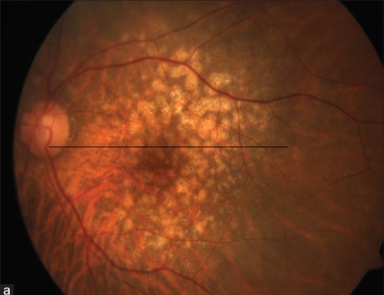
Fundus examination revealed multiple large, yellow-white drusenoid deposits clustered in the posterior pole. Optical coherence tomography (OCT) confirmed the presence of pachydrusen beneath the retinal pigment epithelium (RPE) and demonstrated significant choroidal thickening, with a subfoveal choroidal thickness of 420 µm.
Indocyanine green angiography (ICGA) revealed choroidal vascular hyperpermeability. At the time of examination, there were no signs of choroidal neovascularization (CNV).
The patient was diagnosed with pachychoroid disease and scheduled for close monitoring. Six months later, the patient developed CNV, as evidenced by leakage on fluorescein angiography, and was treated with intravitreal anti-VEGF injections.
Introduction
Pachydrusen is a recently recognized type of drusenoid deposit primarily associated with pachychoroid spectrum diseases.
Unlike the typical drusen seen in age-related macular degeneration (AMD), pachydrusen are linked to thickened choroid and diseases such as central serous chorioretinopathy (CSCR) and polypoidal choroidal vasculopathy (PCV).
Understanding the distinct characteristics of pachydrusen is critical for accurate diagnosis and management of these diseases, as their presence indicates a higher risk of choroidal neovascularization and other complications.
Fundus Explorer Pro
Photograph the retinal findings described here with the phone already in your pocket — 22 D optics and built-in illumination in one handheld unit.
From Choroida — the team behind this siteDisease entity
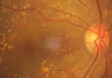
Pachydrusen are sub-RPE deposits larger than 125 µm and are often associated with a thickened choroid. This distinguishes them from the more common soft drusen in eyes with normal or thinner choroids.
Pachydrusen arises from increased choroidal hyperpermeability, a key feature of pachychoroid diseases. The abnormal pachyvessels that characterize these diseases may contribute to the formation of these drusenoid deposits.
Pseudodrusen, on the other hand, are located above the RPE and are associated with thinner choroids. They are often seen in advanced AMD with geographic atrophy.
Epidemiology
Pachydrusen are less commonly seen in patients with non-exudative AMD, occurring in around 11.7% of cases, but are more prevalent in patients with pachychoroid spectrum diseases.
Studies have shown that pachydrusen can be found in up to 49% of cases of CSCR and PCV, particularly in Asian populations where pachychoroid diseases are more common.
Clinical Features
Pachydrusen are large, irregularly shaped deposits found beneath the RPE, primarily in the posterior pole. They differ from soft drusen in their size, shape, and association with a thickened choroid.
On fundoscopic examination, pachydrusen appear as yellow-white lesions clustered in the posterior pole, while OCT imaging confirms their sub-RPE location and reveals accompanying choroidal thickening.
Examination Findings
- Fundoscopy: Pachydrusen appears as large, yellow-white deposits beneath the RPE, usually clustered in the posterior pole.
- OCT Imaging: These deposits are located beneath the RPE, with significant choroidal thickening, differentiating them from soft drusen and pseudodrusen.
- ICGA and Fluorescein Angiography: ICGA shows choroidal hyperpermeability, while fluorescein angiography can reveal leakage if CNV is present.
Differential Diagnosis
- Central serous chorioretinopathy (CSCR)
- Polypoidal choroidal vasculopathy (PCV)
- Age-related macular degeneration (AMD) with soft drusen
- Pseudodrusen
- Peripapillary pachychoroid syndrome
Management
Management of pachydrusen focuses on monitoring for progression to more advanced pachychoroid disease or choroidal neovascularization.
Regular follow-up using OCT and angiography is recommended, as pachydrusen often precedes the development of exudative complications, such as PCV or CNV. Anti-VEGF therapy is indicated if CNV or significant leakage occurs.
Prognosis
Patients with pachydrusen are at increased risk of developing choroidal neovascularization or other exudative complications, leading to potential visual loss if not promptly treated.
Regular monitoring is essential to detect progression early, ensuring timely intervention with therapies like anti-VEGF to preserve vision.


Document what you see
Two smartphone imaging tools built for everyday clinic use — one for the slit lamp, one for the fundus.
From Choroida — the team behind this siteReferences
- Spaide, R. F. (2018). Disease expression in nonexudative age-related macular degeneration varies with choroidal thickness. Retina, 38(4), 708-716.
- Zhang, X., & Sivaprasad, S. (2020). Drusen and pachydrusen: the definition, pathogenesis, and clinical significance. Eye, 1-13.
- Fukuda, Y., Sakurada, Y., Yoneyama, S., Kikushima, W., Sugiyama, A., Matsubara, M., … & Iijima, H. (2019). Clinical and genetic characteristics of pachydrusen in patients with exudative age-related macular degeneration. Scientific Reports, 9(1), 1-7.
- Hosoda, Y., Yoshikawa, M., Miyake, M., Tabara, Y., Ahn, J., Woo, S. J., … & Nagahama Study Group. (2018). CFH and VIPR2 as susceptibility loci in choroidal thickness and pachychoroid disease central serous chorioretinopathy. Proceedings of the National Academy of Sciences, 115(24), 6261-6266.
- Singh, S. R., Oli, A., Mohan, S., Goud, A., Rasheed, M. A., Vupparaboina, K. K., & Chhablani, J. K. (2019). Pachydrusen in Indian population: A hospital-based study. Indian Journal of Ophthalmology, 67(3), 371.
