
HIV retinopathy is a noninfectious microvasculopathy of the retina seen in patients with HIV infection, and immune recovery uveitis is an inflammatory complication that follows immune reconstitution in patients who have had cytomegalovirus retinitis.
The two conditions occur at opposite ends of the immune spectrum, and they both need to be separated from active opportunistic infection.
Antiretroviral therapy has changed the ocular picture of HIV, making sight-threatening opportunistic infection less frequent and inflammatory complications relatively more important.
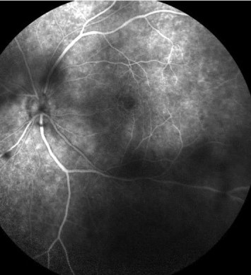
HIV Retinopathy
Definition and Epidemiology
HIV retinopathy, or HIV microvasculopathy, comprises cotton wool spots, intraretinal hemorrhages, and microaneurysms in patients with HIV, without any evidence of opportunistic infection.
It was the most common ocular finding in patients with AIDS before combination antiretroviral therapy became available, seen in roughly half or more of patients in some series.
It is now less frequent and is seen mostly in patients with uncontrolled viremia and low CD4 counts.
Pathogenesis
The mechanism is not fully understood.
Proposed contributors include endothelial damage from HIV and inflammatory cytokines, immune complex deposition, hyperviscosity, and abnormal blood flow.
Clinical Features
Patients are usually asymptomatic, and findings are noted on routine examination.
- Cotton wool spots, typically near the posterior pole and along the arcades
- Dot and blot or flame-shaped hemorrhages
- Microaneurysms and telangiectasia
- Occasional macular edema or capillary nonperfusion
Cotton wool spots resolve over four to six weeks and may recur in other locations.
Visual acuity is usually preserved.
Some patients have subtle deficits in color vision and contrast sensitivity that have been referred to as HIV neuroretinal disorder.
Differential Diagnosis
Cotton wool spots and hemorrhages have many causes:
- Diabetic and hypertensive retinopathy
- Retinal vein occlusion
- Anemia, leukemia, and other blood disorders
- Systemic lupus erythematosus and other vasculitis
- Radiation retinopathy and drug toxicity such as interferon
- Early CMV retinitis, which needs to be excluded (see CMV retinitis)
Large, granular, or hemorrhagic lesions along vessels in a patient with CD4 counts below 50 raise concern for CMV retinitis, and follow-up within days shows expansion of the lesion.
Management
There is no ocular treatment.
Antiretroviral therapy to suppress viral load and restore CD4 counts is the effective intervention.
Patients with low CD4 counts need regular dilated examinations, since CMV retinitis and other opportunistic infections can occur without symptoms (see CMV retinitis management).
Immune Recovery Uveitis
Definition
Immune recovery uveitis (IRU) is intraocular inflammation that develops in patients with HIV and treated CMV retinitis after immune function improves on antiretroviral therapy.
It occurs in eyes in which CMV retinitis is inactive, and the inflammation is a response to residual CMV antigen.
Risk Factors
- CD4 count rising above 100 cells/µL after treatment
- Large area of retinitis at diagnosis
- Involvement of zone 1
- Use of intravitreal cidofovir in some reports
A substantial minority of patients with treated CMV retinitis and immune recovery develop IRU, with a wide range between series.
Clinical Features
Symptoms include floaters, blurred vision, and photophobia.
Findings include:
- Vitritis, which is often prominent
- Anterior chamber inflammation
- Cystoid macular edema
- Epiretinal membrane formation
- Optic disc swelling or neovascularization
- Cataract
- Retinal neovascularization and vitreous hemorrhage in some patients
Vision loss usually comes from macular edema, epiretinal membrane, or cataract.
Management
- Confirm that CMV retinitis is inactive, and exclude other infections such as syphilis and tuberculosis
- Continue antiretroviral therapy and anti-CMV maintenance as advised by the infectious disease team
- Treat anterior inflammation with topical corticosteroids
- Treat macular edema with periocular corticosteroid injection, which has been effective in reported series
- Use oral corticosteroids cautiously and with infectious disease support
- Consider vitrectomy for epiretinal membranes and persistent vitreous opacities
Intravitreal steroids and implants have been used, and the risk of CMV reactivation needs to be considered.
Prognosis
Many patients respond to local steroid therapy, and others have recurrent macular edema.
Prevention depends on early diagnosis of CMV retinitis and on timely antiretroviral therapy.
Fundus Explorer Pro
Photograph the retinal findings described here with the phone already in your pocket — 22 D optics and built-in illumination in one handheld unit.
From Choroida — the team behind this siteScreening and Context
Since the introduction of combination therapy, the incidence of CMV retinitis has fallen sharply in settings where treatment is available, and the disease is now concentrated in patients who present late or who do not take treatment.
In many parts of the world, late presentation remains common, so the ophthalmologist may be the first clinician to diagnose advanced HIV.
Any patient with cotton wool spots, unexplained retinitis, or unexplained uveitis should be offered HIV testing with appropriate counseling.
Practical Points
- Screen patients with CD4 counts below 100 with dilated examination
- Do not assume that cotton wool spots are benign in a patient with a low CD4 count, and re-examine within a short interval
- Keep a low threshold for testing for syphilis, tuberculosis, toxoplasmosis, and PORN in immunosuppressed patients
- Coordinate with the HIV care team on antiretroviral therapy and on drug interactions


Document what you see
Two smartphone imaging tools built for everyday clinic use — one for the slit lamp, one for the fundus.
From Choroida — the team behind this siteReferences
- Holland GN. AIDS and ophthalmology: the first quarter century. Am J Ophthalmol. 2008;145:397-408.
- Freeman WR, Chen A, Henderly DE, et al. Prevalence and significance of acquired immunodeficiency syndrome-related retinal microvasculopathy. Am J Ophthalmol. 1989;107:229-235.
- Jabs DA, Van Natta ML, Holbrook JT, et al. Longitudinal study of the ocular complications of AIDS: 1. Ocular diagnoses at enrollment. Ophthalmology. 2007;114:780-786.
- Karavellas MP, Lowder CY, Macdonald C, Avila CP Jr, Freeman WR. Immune recovery vitritis associated with inactive cytomegalovirus retinitis: a new syndrome. Arch Ophthalmol. 1998;116:169-175.
- Kempen JH, Min YI, Freeman WR, et al. Risk of immune recovery uveitis in patients with AIDS and cytomegalovirus retinitis. Ophthalmology. 2006;113:684-694.
Test yourself
A few questions straight from this article.
-
HIV retinopathy consists of which combination of retinal findings?
HIV retinopathy, or HIV microvasculopathy, comprises cotton wool spots, intraretinal haemorrhages and microaneurysms in a patient with HIV and no evidence of opportunistic infection. -
Which process is proposed to cause HIV retinopathy?
The mechanism is not fully understood. Proposed contributors include endothelial damage from HIV and inflammatory cytokines, immune complex deposition, hyperviscosity and abnormal blood flow. -
Over what period do cotton wool spots in HIV retinopathy resolve?
Cotton wool spots resolve over four to six weeks and may recur in other locations. Patients are usually asymptomatic and visual acuity is generally preserved. -
Which feature in an HIV patient with retinal lesions raises concern for CMV retinitis rather than HIV retinopathy?
Large, granular or haemorrhagic lesions along vessels in a patient with a CD4 count below 50 raise concern for CMV retinitis, and follow-up within days shows the lesion expanding. -
What is the effective treatment for HIV retinopathy?
There is no ocular treatment for HIV retinopathy. Antiretroviral therapy to suppress viral load and restore CD4 counts is the effective intervention, alongside regular dilated examination when CD4 counts are low. -
Immune recovery uveitis develops under which circumstances?
Immune recovery uveitis is intraocular inflammation developing after immune function improves on antiretroviral therapy, in eyes where CMV retinitis is inactive. The inflammation is a response to residual CMV antigen. -
Which factor increases the risk of immune recovery uveitis?
Risk factors include a CD4 count rising above 100 cells per microlitre after treatment, a large area of retinitis at diagnosis, zone 1 involvement and, in some reports, intravitreal cidofovir. -
How is macular oedema from immune recovery uveitis treated?
Macular oedema is treated with periocular corticosteroid injection, which has been effective in reported series. Anterior inflammation is treated topically, and vitrectomy is reserved for epiretinal membranes and persistent vitreous opacities. -
Which presentation should prompt an offer of HIV testing with appropriate counselling?
Any patient with cotton wool spots, unexplained retinitis or unexplained uveitis should be offered HIV testing with appropriate counselling, since the ophthalmologist may be the first clinician to diagnose advanced HIV. -
What is the course of immune recovery uveitis after treatment is started?
Many patients respond to local steroid therapy while others have recurrent macular oedema. Prevention depends on early diagnosis of CMV retinitis and timely antiretroviral therapy.

Writes and reviews the clinical articles on Educate. Every post is checked against peer-reviewed sources before it goes live.


