
Uveal effusion syndrome produces choroidal detachment, often with an associated non-rhegmatogenous retinal detachment, in an eye without a retinal break, an inflammatory cause, or hypotony, and it is most classically seen in a nanophthalmic eye with an abnormally thick sclera that impedes the normal drainage of fluid from the choroid.
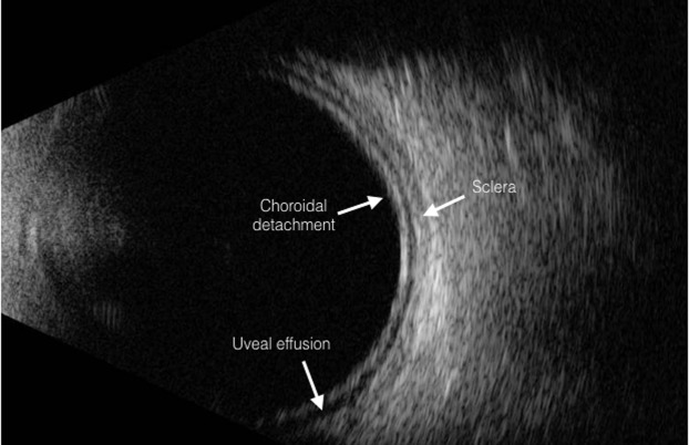
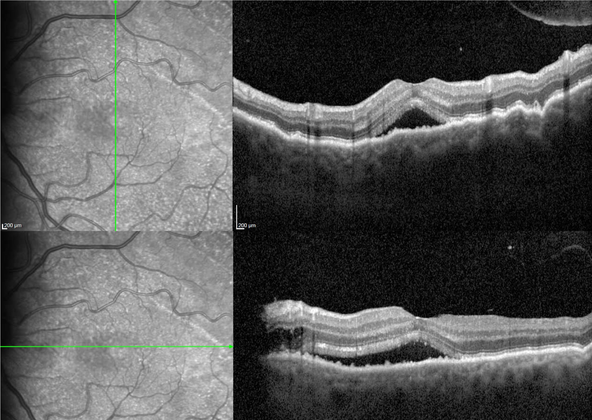
Recognizing that the sclera, not the choroid or retina, is often the actual problem is what makes the surgical treatment for this condition make sense.
The Role of Scleral Thickness
Fluid normally exits the choroid through the vortex veins and through the sclera’s own permeability, and in uveal effusion syndrome an abnormally thick, sometimes structurally abnormal sclera impedes this outflow, either by compressing the vortex veins as they pass through the scleral canals or by reducing the sclera’s own transscleral fluid permeability.
The result is progressive accumulation of fluid in the suprachoroidal and, in more advanced cases, subretinal space, without any of the usual causes such as a retinal break, inflammation, or ocular hypotony.
Classification
Uveal effusion syndrome is typically divided by cause.
- Nanophthalmic (type 1): occurring in a small eye with a short axial length, thick sclera, and often high hyperopia, the classic setting for the condition (see nanophthalmos)
- Non-nanophthalmic with abnormal sclera (type 2): a normal-sized eye but with abnormally thick or structurally abnormal sclera, sometimes associated with systemic connective tissue conditions
- Idiopathic (type 3): occurring in an eye with normal axial length and no clearly identified scleral abnormality, a diagnosis of exclusion
Fundus Explorer Pro
Photograph the retinal findings described here with the phone already in your pocket — 22 D optics and built-in illumination in one handheld unit.
From Choroida — the team behind this siteClinical Features
- Slowly progressive, painless visual loss and visual field changes
- Bullous, shifting choroidal detachment, often with an associated serous retinal detachment that follows gravity and changes with head position, unlike a fixed rhegmatogenous detachment
- A characteristic leopard-spot pattern of pigmentary mottling in the fundus periphery, seen in chronic cases from long-standing subretinal fluid
- High hyperopia and a short, crowded anterior segment in the nanophthalmic form
- Absence of a retinal break on careful examination, distinguishing it from rhegmatogenous detachment
Diagnosis
- B-scan ultrasonography shows thickened sclera and choroid, along with the characteristic smooth, shifting choroidal detachment
- Axial length measurement, which is short in nanophthalmic cases
- Careful examination for a retinal break, essential to exclude a rhegmatogenous cause before attributing the detachment to uveal effusion
- Systemic evaluation for connective tissue or other disease in non-nanophthalmic cases, since scleral abnormality can occasionally be part of a broader systemic condition
Differential Diagnosis
- Rhegmatogenous retinal detachment, excluded by the absence of a retinal break and by the smooth, shifting nature of the effusion
- Posterior scleritis, which typically has pain and an inflammatory component, along with a different ultrasonographic appearance (see posterior scleritis)
- Choroidal detachment from hypotony after surgery or trauma, distinguished by the clinical context and by intraocular pressure findings
- Choroidal tumor, excluded by the diffuse rather than focal nature of the effusion and by imaging characteristics
Management
Observation
Mild cases with stable, limited effusion and preserved vision may be observed, since the condition can remain stable over long periods in some patients.
Scleral Surgery
Scleral window surgery, removing partial-thickness patches of sclera, usually over the vortex veins, is the definitive treatment for nanophthalmic and thick-sclera cases, aiming to relieve the outflow obstruction directly, and it can be combined with sclerectomy to further thin the surrounding sclera.
Some surgeons add vortex vein decompression as part of the same procedure.
Management of Coexisting Findings
Nanophthalmic eyes are also predisposed to angle closure glaucoma and require attention to this risk separately from the effusion itself, and cataract or other intraocular surgery in these eyes carries a higher risk of intraoperative and postoperative choroidal effusion, requiring specific precautions.
Prognosis
Scleral surgery improves outcomes substantially in appropriately selected cases, particularly nanophthalmic eyes, with resolution or significant improvement of the effusion in most patients.
Untreated, progressive disease can lead to significant, sometimes severe, visual loss, which is why recognizing the condition and its underlying scleral basis matters for offering effective treatment rather than continued observation alone.


Document what you see
Two smartphone imaging tools built for everyday clinic use — one for the slit lamp, one for the fundus.
From Choroida — the team behind this siteReferences
- Uyama M, Takahashi K, Kozaki J, et al. Uveal effusion syndrome: clinical features, surgical treatment, histologic examination of the sclera, and pathophysiology. Ophthalmology. 2000;107:441-449.
- Gass JD, Jallow S. Idiopathic serous detachment of the choroid, ciliary body, and retina (uveal effusion syndrome). Ophthalmology. 1982;89:1018-1032.
- Jackson TL, Hussain A, Morley AM, et al. Scleral hydraulic conductivity and macromolecular diffusion in patients with uveal effusion syndrome. Invest Ophthalmol Vis Sci. 2008;49:5033-5040.
- Brockhurst RJ. Nanophthalmos with uveal effusion: a new clinical entity. Arch Ophthalmol. 1975;93:1289-1299.
