
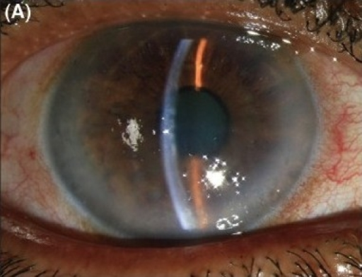
Fuchs endothelial dystrophy is the most common primary corneal endothelial disease, typically bilateral, slowly progressive, and more frequent in women, and genetic research over the past two decades has identified a specific repeat expansion in the TCF4 gene as the leading cause in a large proportion of cases, reframing a condition once considered purely age-related as one with a strong, identifiable hereditary basis in many patients.
Clinical Course
Fuchs dystrophy progresses through recognizable stages, though the pace varies considerably between patients.
- Early disease: scattered guttae, small excrescences of abnormal Descemet membrane visible as a beaten-metal appearance on the corneal endothelium, often asymptomatic and discovered incidentally
- Progressive guttae formation and endothelial cell loss, with gradually reduced endothelial function
- Stromal edema: once endothelial pump function falls below what is needed, corneal thickening develops, causing blurred vision, classically worse on waking
- Epithelial and subepithelial changes: painful bullae, and eventually subepithelial fibrosis in advanced, longstanding disease
Genetics
A trinucleotide (CTG) repeat expansion in the TCF4 gene is the most common identified genetic cause of Fuchs endothelial dystrophy, present in a majority of patients in several population studies, particularly those of European ancestry, though the strength of this association varies somewhat between populations.
Other genes have been implicated in a smaller proportion of cases, including some early-onset and familial forms with mutations in genes such as COL8A2, though these represent a minority of overall disease compared with the TCF4 repeat expansion.
The genetic basis explains the strong familial clustering long observed in Fuchs dystrophy and raises the future possibility of genetic testing to identify at-risk relatives or to stratify patients for emerging non-surgical treatments targeting the underlying repeat expansion mechanism.
All-fit Slit-Lamp Adapter
Record and share exactly what you see at the slit lamp. One adapter fits any slit lamp or surgical microscope — and any smartphone.
From Choroida — the team behind this siteDiagnosis
- Slit-lamp examination showing the characteristic guttae and beaten-metal endothelial appearance, best appreciated with specular reflection technique
- Specular or confocal microscopy to quantify endothelial cell density and characterize guttae
- Corneal pachymetry, since increasing central corneal thickness reflects declining endothelial function and helps track disease progression over time
- Anterior segment OCT, useful for measuring corneal thickness and characterizing Descemet membrane changes, particularly helpful when the view is limited by stromal or epithelial edema
Impact on Cataract Surgery Planning
Fuchs dystrophy has particular relevance to cataract surgery planning, since phacoemulsification imposes additional stress on an endothelium that may already have reduced reserve.
- Preoperative specular microscopy and pachymetry help estimate endothelial reserve and the risk of postoperative decompensation
- Patients with more advanced guttae or reduced cell counts may be candidates for combined cataract surgery with endothelial keratoplasty (a “triple procedure”) rather than cataract surgery alone, anticipating that the endothelium would decompensate from cataract surgery stress even if it was managing acceptably beforehand
- Surgical technique modifications, including gentler phacoemulsification parameters and use of a dispersive viscoelastic, are used to minimize additional endothelial trauma in eyes with compromised endothelial reserve
Management
Early Disease
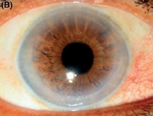
Hypertonic saline and other measures used for general endothelial decompensation provide symptomatic relief once mild edema develops, without altering the underlying disease course (see corneal endothelial decompensation).
Surgical Treatment
Once vision or comfort is significantly affected by stromal edema, endothelial keratoplasty, most often DMEK given its excellent visual outcomes in Fuchs dystrophy specifically, replaces the diseased endothelium (see DSEK/DMEK).
Emerging and Investigational Approaches
Descemet stripping only, without a donor graft, relies on migration and proliferation of the patient’s own peripheral endothelial cells to repopulate the central cornea and has shown promising results in carefully selected patients with adequate peripheral endothelial reserve, avoiding the need for donor tissue in this select group.
Pharmacologic approaches to stimulate endothelial cell proliferation, including Rho-kinase inhibitors, are under active investigation as potential future adjuncts or alternatives to transplantation.
Prognosis
Fuchs dystrophy progresses slowly over years to decades in most patients, and many never require surgical intervention during their lifetime.
For those who do progress to visually significant disease, modern endothelial keratoplasty offers excellent outcomes, with most patients achieving good vision and faster recovery than was possible with older full-thickness transplant techniques.


Document what you see
Two smartphone imaging tools built for everyday clinic use — one for the slit lamp, one for the fundus.
From Choroida — the team behind this siteReferences
- Wieben ED, Aleff RA, Tosakulwong N, et al. A common trinucleotide repeat expansion within the transcription factor 4 (TCF4) gene predicts Fuchs corneal dystrophy. PLoS One. 2012;7:e49083.
- Eghrari AO, Riazuddin SA, Gottsch JD. Fuchs corneal dystrophy. Prog Mol Biol Transl Sci. 2015;134:79-97.
- Krachmer JH, Purcell JJ Jr, Young CW, Bucher KD. Corneal endothelial dystrophy: a study of 64 families. Arch Ophthalmol. 1978;96:2036-2039.
- Borkar DS, Veldman P, Colby KA. Treatment of Fuchs endothelial dystrophy by Descemet stripping without endothelial keratoplasty. Cornea. 2016;35:1267-1273.
Test yourself
A few questions straight from this article.
-
Which description fits Fuchs endothelial dystrophy?
Fuchs endothelial dystrophy is the most common primary corneal endothelial disease, typically bilateral, slowly progressive, and more frequent in women. -
What are the guttae of Fuchs endothelial dystrophy?
Guttae are small excrescences of abnormal Descemet membrane, seen as a beaten-metal appearance on the corneal endothelium, and are often an incidental finding early on. -
Which genetic change is the commonest identified cause of Fuchs endothelial dystrophy?
A trinucleotide CTG repeat expansion in the TCF4 gene is the most common identified genetic cause, present in a majority of patients in several population studies, particularly those of European ancestry. -
Mutations in COL8A2 account for which part of Fuchs endothelial dystrophy?
Genes such as COL8A2 are implicated in a smaller proportion of cases, including some early-onset and familial forms, a minority compared with the TCF4 repeat expansion. -
Which slit-lamp technique best demonstrates the guttae of Fuchs dystrophy?
The characteristic guttae and beaten-metal endothelial appearance are best appreciated at the slit lamp using the specular reflection technique. -
Which patients with Fuchs dystrophy may be offered combined cataract surgery and an endothelial graft?
Patients with more advanced guttae or reduced cell counts may be candidates for a triple procedure, anticipating that the endothelium would decompensate from the stress of cataract surgery alone. -
Which modifications reduce endothelial trauma during cataract surgery in Fuchs dystrophy?
Gentler phacoemulsification parameters and the use of a dispersive viscoelastic are used to minimise additional endothelial trauma in eyes with compromised endothelial reserve. -
What does Descemet stripping only rely on to clear the cornea in Fuchs dystrophy?
Descemet stripping without a donor graft relies on migration and proliferation of the patient's own peripheral endothelial cells, so it suits selected patients with adequate peripheral endothelial reserve. -
Which drug class is under investigation to stimulate corneal endothelial cell proliferation?
Pharmacologic approaches to stimulate endothelial cell proliferation, including Rho-kinase inhibitors, are under active investigation as future adjuncts or alternatives to transplantation. -
What is the usual natural course of Fuchs endothelial dystrophy?
Fuchs dystrophy progresses slowly over years to decades in most patients, and many never require surgical intervention during their lifetime.

Writes and reviews the clinical articles on Educate. Every post is checked against peer-reviewed sources before it goes live.


