Laser peripheral iridotomy creates a small, full-thickness opening in the peripheral iris using a Nd:YAG laser, giving aqueous humor an alternate forward path from the posterior to the anterior chamber and relieving pupillary block, the mechanism underlying acute angle-closure glaucoma, discussed in its own dedicated article on this site.
It is one of the most frequently performed laser procedures in ophthalmology, used both to treat acute angle closure emergently and, just as often, to prevent a first episode in an eye identified as anatomically at risk before any acute event occurs.
Understanding exactly what the iridotomy does and does not fix clarifies why it remains highly effective for pupillary block specifically, but offers little to no benefit in angle closure driven by other mechanisms, including the malignant glaucoma discussed in its own dedicated article on this site.
The procedure’s brevity and generally low complication rate are part of why it has become such a routine, high-volume part of everyday glaucoma practice worldwide.
Mechanism
Pupillary block occurs when the flow of aqueous from the posterior to the anterior chamber through the pupil is impeded by apposition between the iris and the lens, causing pressure to build in the posterior chamber and push the peripheral iris forward against the trabecular meshwork.
A peripheral iridotomy creates a direct, alternate opening between the posterior and anterior chambers, equalizing pressure across the iris and eliminating the forward bowing that was crowding and potentially closing the angle.
Because this mechanism is specific to pupillary block, an iridotomy does nothing to relieve angle closure from other causes, such as plateau iris configuration or the posterior pushing mechanism of malignant glaucoma, which is exactly why a patent iridotomy that fails to resolve elevated pressure should immediately raise suspicion for one of these alternate mechanisms.
Technique
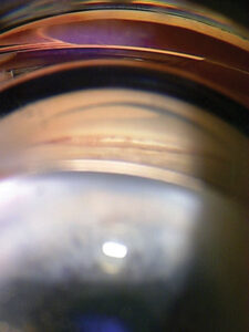
The procedure is performed at a slit lamp using a specialized iridotomy contact lens to focus the laser accurately, typically targeting a peripheral, superior location on the iris that will be naturally covered by the upper eyelid, minimizing visual side effects such as glare or a new linear image artifact.
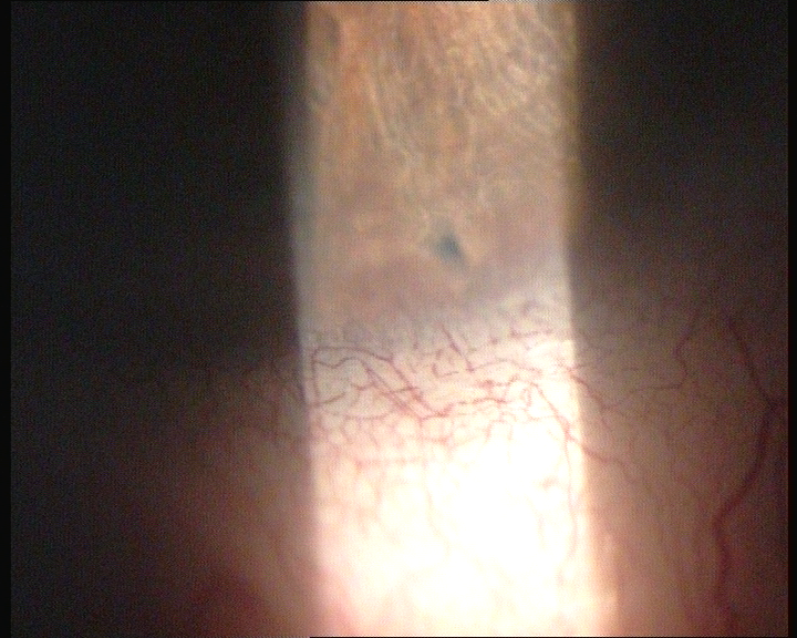
A Nd:YAG laser delivers short pulses of energy to photodisrupt the iris tissue, and the surgeon confirms the opening is full-thickness by visualizing a plume of pigment or an actual view through to the underlying lens capsule.
Topical anesthesia alone is sufficient, and the procedure typically takes only a few minutes, with the patient generally able to resume normal activities the same day.
Confirming true patency, rather than assuming success based on the laser response alone, matters enough that many surgeons deliberately check for a transilluminated opening or a clear view to the lens before ending the session.
All-fit Slit-Lamp Adapter
Record and share exactly what you see at the slit lamp. One adapter fits any slit lamp or surgical microscope — and any smartphone.
From Choroida — the team behind this siteIndications
- Acute angle-closure glaucoma, once the acute attack has been broken sufficiently to allow adequate visualization and laser access to the iris
- Primary angle closure suspects, eyes identified on gonioscopy as anatomically narrow and at risk, treated prophylactically before any acute event occurs
- Fellow eyes of a patient who has already had an acute angle-closure attack, given the strong likelihood of similarly predisposed anatomy in the second eye
- Chronic angle closure with a component of pupillary block contributing to progressive angle narrowing over time
Complications
Transient intraocular pressure elevation is common in the hours following the procedure, and pressure is typically checked before the patient leaves, with a short course of topical medication used if elevated.
Glare, halos, or a new linear visual artifact can occur, particularly when the iridotomy is not adequately covered by the upper eyelid, which is part of why careful attention to placement location matters during the procedure itself.
A small amount of bleeding at the treatment site is common and generally self-limited, and localized lens capsule changes at the treatment site are occasionally seen but rarely visually significant.
When It Does Not Work
A confirmed patent iridotomy that fails to relieve elevated pressure or a persistently shallow anterior chamber should prompt reconsideration of the diagnosis, specifically raising the possibility of malignant glaucoma, plateau iris syndrome, or another non-pupillary-block mechanism of angle closure.
This is precisely the clinical scenario where confusing malignant glaucoma with ordinary pupillary block, discussed in detail in the malignant glaucoma article on this site, becomes most dangerous, since further attempts at pupillary-block-directed treatment do nothing to address a fundamentally different underlying mechanism.
Recognizing this failure pattern quickly, rather than assuming the iridotomy simply needs more time to work, is what actually protects the patient from a delayed diagnosis of a genuinely different and more urgent condition.


Document what you see
Two smartphone imaging tools built for everyday clinic use — one for the slit lamp, one for the fundus.
From Choroida — the team behind this siteReferences
- American Academy of Ophthalmology. Basic and Clinical Science Course, Section 10: Glaucoma.
- Vera V, Naqi A, Belovay GW, et al. Dysphotopsia after temporal versus superior laser peripheral iridotomy: a prospective randomized paired eye trial. American Journal of Ophthalmology.
- He M, Friedman DS, Ge J, et al. Laser peripheral iridotomy in primary angle-closure suspects: biometric and gonioscopic outcomes. Ophthalmology.
Test yourself
A few questions straight from this article.
-
Laser peripheral iridotomy relieves pupillary block by creating what?
A Nd:YAG laser creates a small full-thickness opening in the peripheral iris, giving aqueous an alternate forward path from the posterior to the anterior chamber. -
Pupillary block develops when aqueous flow through the pupil is impeded by which apposition?
In pupillary block, apposition between the iris and the lens impedes aqueous passage from the posterior to the anterior chamber through the pupil. -
In pupillary block, rising posterior chamber pressure produces which anatomical change?
Pressure building in the posterior chamber pushes the peripheral iris forward against the trabecular meshwork, crowding and potentially closing the angle. -
How does a patent iridotomy abolish the forward bowing of the iris?
The iridotomy provides a direct alternate opening between the two chambers, equalising pressure across the iris and eliminating the forward bowing that crowded the angle. -
What confirms during the procedure that a laser iridotomy is full-thickness?
The surgeon confirms a full-thickness opening by seeing a plume of pigment or an actual view through to the underlying lens capsule, rather than assuming success from the laser response. -
What anaesthesia does laser peripheral iridotomy require?
The procedure is done at the slit lamp under topical anaesthesia alone, takes only a few minutes, and the patient can usually resume normal activities the same day. -
Which eye receives a prophylactic iridotomy before any acute attack has occurred?
Primary angle closure suspects, identified on gonioscopy as anatomically narrow and at risk, are treated prophylactically before any acute event occurs. -
Why is the fellow eye treated after an acute angle-closure attack in one eye?
Fellow eyes of a patient who has had an acute angle-closure attack are treated because of the strong likelihood of similarly predisposed anatomy in the second eye. -
Which complication is checked for before the patient leaves after laser peripheral iridotomy?
Transient pressure elevation is common in the hours after iridotomy, so pressure is typically checked before the patient leaves and treated with a short course of topical medication if raised. -
A patent iridotomy with persistently high pressure and a shallow chamber should prompt which consideration?
An iridotomy addresses pupillary block only, so a confirmed patent opening that fails to relieve pressure should raise suspicion of malignant glaucoma or another non-pupillary-block mechanism.

Writes and reviews the clinical articles on Educate. Every post is checked against peer-reviewed sources before it goes live.