
Traumatic macular hole is a full-thickness foveal defect that develops after blunt or penetrating injury to the eye.
It is typically seen in young men after sports injuries, fights, or projectile trauma, and it differs from an idiopathic hole because a healthy vitreous is often involved and the hole can close by itself.
That difference shapes management, since a period of observation is often reasonable before considering surgery.
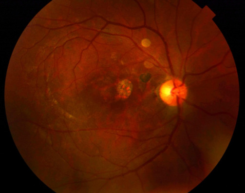
Mechanism of Traumatic Macular Hole
Blunt trauma compresses the globe in the anteroposterior direction and expands it at the equator.
The resulting shock wave and sudden change in shape stress the fovea, which is the thinnest part of the retina.
Several mechanisms have been proposed:
- Contusion of the retina, with outer retinal necrosis and cystic degeneration
- Acute vitreous traction on the fovea at the moment of impact
- Tangential traction from an epiretinal membrane that forms afterward
- Coup and contrecoup forces that produce a direct macular rupture
Holes may form immediately or appear days to months after the injury, when commotio retinae has resolved and the underlying damage becomes visible (see commotio retinae).
Traumatic macular hole is reported in a small percentage of patients with closed globe contusion.
Clinical Presentation
Patients complain of sudden central vision loss, with a central scotoma and distortion.
Visual acuity varies widely, and it is often markedly reduced at presentation.
On examination the hole appears as a round red foveal lesion with or without a cuff of subretinal fluid.
Associated findings from the same injury are common:
- Commotio retinae
- Choroidal rupture, which is often concentric to the disc (see choroidal rupture)
- Retinal hemorrhage
- Vitreous hemorrhage
- Peripheral retinal tears and dialyses
- Optic nerve injury
- Hyphema, angle recession, and lens injury
A full dilated examination with scleral depression is therefore needed, since additional injuries determine prognosis.
Fundus Explorer Pro
Photograph the retinal findings described here with the phone already in your pocket — 22 D optics and built-in illumination in one handheld unit.
From Choroida — the team behind this siteImaging
OCT confirms the diagnosis and gives measurements of the hole.
Findings include a full-thickness defect with rounded or irregular edges, intraretinal cysts, subretinal fluid, and sometimes a lamellar component.
The presence of an operculum or vitreous attachment helps decide whether surgery is likely to be needed.
Fluorescein angiography shows a window defect at the hole and may show choroidal rupture or CNV.
Autofluorescence helps assess RPE damage.
Repeat OCT during observation shows whether the hole is closing, and closure is seen as fusion of the edges with restoration of the foveal contour.
Differential Diagnosis
Not every foveal defect after trauma is a full-thickness macular hole.
- Lamellar hole or cystic foveal degeneration, which keeps an intact outer retinal floor
- Pseudohole from an epiretinal membrane
- Commotio retinae with foveal disruption of the outer bands but an intact inner retina
- Choroidal rupture with overlying retinal and RPE damage
- Solar or laser injury, in which the history and pattern differ
OCT resolves most of these questions, and it should be part of every examination after ocular contusion when acuity is worse than the external findings explain.
Spontaneous Closure
Traumatic macular holes can close spontaneously, unlike most idiopathic holes.
Published series report closure in roughly a substantial minority of cases, and some report closure in about half.
Factors that favor spontaneous closure are:
- Younger age
- Smaller hole diameter
- Absence of a large cuff of subretinal fluid
- Absence of tractional membranes
- Shorter time since injury
Closure usually happens within the first three to six months, and a longer observation period is not usually helpful.
Spontaneous closure is often accompanied by improved acuity, although the final result depends on foveal outer retinal recovery.
Management
Observation
For small, recent holes in young patients, observation with serial OCT for three months or longer is a reasonable first step.
Follow-up visits monitor hole size and the vitreoretinal interface.
Patients should be counseled on the possibility of spontaneous closure and on the option of surgery if the hole persists.
Surgery
Pars plana vitrectomy with ILM peeling and gas tamponade is the standard surgical approach (see pars plana vitrectomy).
Surgery is considered when:
- The hole is large or enlarging
- There is a persistent hole after observation
- There is a retinal detachment or tractional membrane
- There is vitreous hemorrhage or another indication for vitrectomy
Series report closure rates of around 90% or more after vitrectomy with ILM peeling, though visual outcome is less predictable than in idiopathic holes because of damage to the RPE and photoreceptors.
Postoperative face-down positioning is used in many centers, although evidence for its necessity is limited in smaller holes.
Surgery in young patients requires attention to cataract formation and to management of fellow-eye protection.
See also the general staging and surgical principles of macular hole (see full-thickness macular hole).
Related Injuries
Choroidal rupture and macular RPE damage limit the visual potential and can cause CNV.
Protective eyewear is the main preventive measure for sports and work.
Prognosis
Visual outcome is variable.
Eyes that close spontaneously or after surgery often improve, although many have residual reduction from foveal damage.
Poor prognostic signs include large holes, choroidal rupture through the fovea, macular pigment epithelial damage, and delayed treatment.
Patients should be followed for CNV, epiretinal membrane, and retinal detachment.


Document what you see
Two smartphone imaging tools built for everyday clinic use — one for the slit lamp, one for the fundus.
From Choroida — the team behind this siteReferences
- Johnson RN, McDonald HR, Lewis H, et al. Traumatic macular hole: observations, pathogenesis, and results of vitrectomy surgery. Ophthalmology. 2001;108:853-857.
- Miller JB, Yonekawa Y, Eliott D, et al. Long-term follow-up and outcomes in traumatic macular holes. Am J Ophthalmol. 2015;160:1255-1258.
- Yamashita T, Uemara A, Uchino E, Doi N, Ohba N. Spontaneous closure of traumatic macular hole. Am J Ophthalmol. 2002;133:230-235.
- Amari F, Ogino N, Matsumura M, Negi A, Honda Y. Vitreous surgery for traumatic macular holes. Retina. 1999;19:410-413.
- Duker JS, Kaiser PK, Binder S, et al. The International Vitreomacular Traction Study Group classification of vitreomacular adhesion, traction, and macular hole. Ophthalmology. 2013;120:2611-2619.
Test yourself
A few questions straight from this article.
-
What defines a traumatic macular hole?
Traumatic macular hole is a full-thickness foveal defect developing after blunt or penetrating injury, seen typically in young men after sports injuries, fights or projectile trauma. -
How does blunt trauma deform the globe in a way that produces a macular hole?
Blunt trauma compresses the globe anteroposteriorly and expands it at the equator. The resulting shock wave and change in shape stress the fovea, the thinnest part of the retina. -
How does traumatic macular hole differ from an idiopathic macular hole?
Unlike most idiopathic holes, a traumatic hole often involves a healthy vitreous and can close spontaneously. That difference is what makes a period of observation reasonable. -
What is the typical fundus appearance of a traumatic macular hole?
On examination the hole appears as a round red foveal lesion with or without a cuff of subretinal fluid. A full dilated examination with scleral depression is needed because associated injuries determine prognosis. -
Which OCT feature identifies a lamellar hole rather than a full-thickness traumatic macular hole?
A lamellar hole or cystic foveal degeneration keeps an intact outer retinal floor, whereas a traumatic macular hole is a full-thickness defect. OCT resolves most post-contusion foveal questions. -
Which factor favours spontaneous closure of a traumatic macular hole?
Younger age, smaller hole diameter, absence of a large subretinal fluid cuff, absence of tractional membranes and a shorter time since injury all favour spontaneous closure. -
What is a reasonable first step for a small, recent traumatic macular hole in a young patient?
Observation with serial OCT for three months or longer is reasonable for small recent holes in young patients, since spontaneous closure usually happens within the first three to six months. -
Which situation calls for vitrectomy rather than continued observation of a traumatic macular hole?
Surgery is considered for a large or enlarging hole, a hole persisting after observation, retinal detachment or tractional membrane, or another indication for vitrectomy such as vitreous haemorrhage. -
Which operation is standard for a persistent traumatic macular hole, and what closure rate is reported?
Pars plana vitrectomy with ILM peeling and gas tamponade is standard, with reported closure rates around 90 percent or more. Visual outcome is less predictable than in idiopathic holes because of RPE and photoreceptor damage. -
Which finding predicts a poor visual outcome in traumatic macular hole?
Poor prognostic signs include large holes, choroidal rupture through the fovea, macular pigment epithelial damage and delayed treatment. Patients also need follow-up for CNV, epiretinal membrane and retinal detachment.

Writes and reviews the clinical articles on Educate. Every post is checked against peer-reviewed sources before it goes live.


