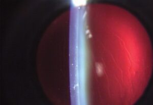
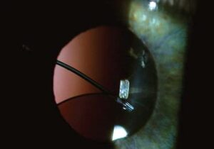
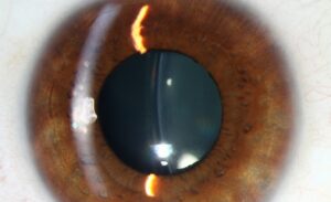
Scleral-fixated intraocular lens implantation secures an IOL directly to the sclera with sutures or, in newer techniques, without sutures at all, providing stable lens fixation in eyes that lack adequate capsular support for a conventional in-the-bag or sulcus-placed lens.
It represents one of several options for managing an eye without sufficient capsular support, alongside anterior chamber lenses and iris-fixated lenses, and the choice among these alternatives depends heavily on the specific anatomy and circumstances of the individual eye.
Understanding why capsular support matters so much, and what happens when it is absent or severely compromised, explains why this more technically demanding fixation technique exists as a distinct category of intraocular lens surgery at all.
These cases represent a comparatively small but consistently encountered subset of intraocular lens surgery, and they demand a level of surgical planning and technique well beyond that of routine, uncomplicated cataract surgery.
Why Capsular Support Matters
A standard intraocular lens relies on an intact capsular bag, or at least adequate residual capsular and zonular support, to hold it securely and stably centered within the eye, following the same anatomic principle used in routine phacoemulsification, discussed in its own dedicated article on this site.
When capsular support is inadequate, whether from a complicated cataract surgery with significant capsular rupture, zonular dehiscence from trauma or a connective tissue disorder, or the need to remove a previously dislocated lens entirely, a standard in-the-bag lens cannot be placed safely.
In these situations, the lens needs an alternate fixation point, and scleral fixation uses the sclera itself, a structurally robust and stable tissue, as that alternate anchor.
Iris-fixated lenses and anterior chamber lenses offer alternate solutions to the same underlying problem, and the choice among all these options depends on factors including iris integrity, angle anatomy, and surgeon experience and preference.
Techniques
- Sutured scleral fixation, using permanent sutures passed through the sclera to secure specialized haptics or eyelets on the intraocular lens, the traditional and longest-established approach
- Sutureless scleral fixation techniques, including haptic externalization methods where the lens haptics are threaded through scleral tunnels and secured without any suture material, avoiding suture-related long-term complications discussed below
- Glued intrascleral fixation, using fibrin glue to secure externalized haptics within scleral flaps, another suture-free approach that has gained popularity in some practices
All-fit Slit-Lamp Adapter
Record and share exactly what you see at the slit lamp. One adapter fits any slit lamp or surgical microscope — and any smartphone.
From Choroida — the team behind this siteWhy Sutureless Techniques Have Gained Favor
Sutures used for scleral fixation can degrade, break, or erode through the overlying conjunctiva over time, sometimes years after the original surgery, occasionally causing lens dislocation, exposure, or an increased risk of endophthalmitis at the site of suture erosion.
Sutureless techniques avoid this specific long-term suture-related failure mode entirely, since there is no suture material present to degrade or erode in the first place.
This durability advantage is a major reason sutureless approaches have gained considerable favor among surgeons who regularly perform this type of fixation, even though sutured techniques remain a well-established, effective option, particularly in surgeons and centers most experienced with that specific approach.
Indications
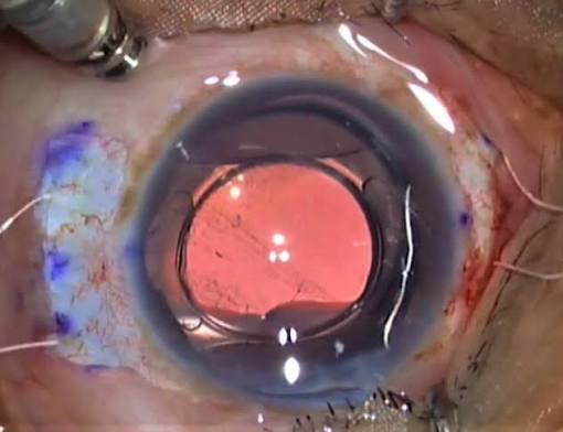
Aphakia without adequate capsular support, whether from complicated cataract surgery, trauma, or removal of a previously dislocated lens, is the core indication for this technique.
Ectopia lentis from connective tissue disorders such as Marfan syndrome, discussed in its own dedicated article on this site, and Weill-Marchesani syndrome, also discussed in its own dedicated article on this site, can require scleral-fixated lens implantation when the native zonular support is too compromised for any lens to rely on it.
Dislocated intraocular lenses requiring exchange, where the original fixation has failed and adequate capsular support for a standard replacement is no longer present, are another common scenario prompting this approach.
Complications
Lens tilt or decentration can occur if scleral fixation is not achieved symmetrically or securely, affecting visual quality even when the lens itself remains stable and well retained overall.
Suture-related complications, discussed above, are specific to sutured technique, while haptic erosion or exposure is the corresponding technique-specific risk more associated with sutureless approaches.
Vitreous hemorrhage, retinal detachment, and cystoid macular edema are recognized general risks given the posterior segment manipulation often involved in these more complex cases, particularly when combined with vitrectomy to address associated vitreous prolapse or other posterior segment issues.
Given this complexity, careful preoperative planning and, where possible, referral to a surgeon specifically experienced with these techniques meaningfully improves the likelihood of a stable, well-centered long-term result.


Document what you see
Two smartphone imaging tools built for everyday clinic use — one for the slit lamp, one for the fundus.
From Choroida — the team behind this siteReferences
- Agarwal A, Kumar DA, Jacob S, et al. Fibrin glue-assisted sutureless posterior chamber intraocular lens fixation in eyes with deficient posterior capsules. Journal of Cataract and Refractive Surgery.
- American Academy of Ophthalmology. Basic and Clinical Science Course, Section 11: Lens and Cataract.
- Yamane S, Sato S, Maruyama-Inoue M, Kadonosono K. Flanged intrascleral intraocular lens fixation with double-needle technique. Ophthalmology.
Test yourself
A few questions straight from this article.
-
Scleral-fixated intraocular lens implantation is chosen for an eye with which deficiency?
Scleral fixation provides stable lens fixation in eyes that lack adequate capsular support for a conventional in-the-bag or sulcus-placed intraocular lens. -
Which tissue serves as the alternate anchor in scleral fixation of an intraocular lens?
When capsular support is inadequate the lens needs an alternate fixation point, and scleral fixation uses the sclera, a structurally robust and stable tissue, as that anchor. -
A conventional in-the-bag intraocular lens depends on which support to stay centred?
A standard lens relies on an intact capsular bag, or at least adequate residual capsular and zonular support, to hold it securely and stably centred in the eye. -
Which situation leaves an eye without adequate capsular support for a standard lens?
Zonular dehiscence from trauma or a connective tissue disorder, significant capsular rupture, or removal of a dislocated lens all leave support inadequate for an in-the-bag lens. -
How does traditional sutured scleral fixation secure the intraocular lens?
Sutured scleral fixation, the traditional and longest-established approach, passes permanent sutures through the sclera to secure specialised haptics or eyelets on the lens. -
Glued intrascleral fixation secures externalised haptics using which material?
Glued intrascleral fixation uses fibrin glue to secure externalised haptics within scleral flaps, a suture-free approach that has gained popularity in some practices. -
Which long-term failure mode affects sutured scleral fixation years after surgery?
Fixation sutures can degrade, break or erode through the overlying conjunctiva, sometimes years after the original surgery, which is the failure mode sutureless methods avoid. -
Suture erosion through the conjunctiva after scleral fixation raises the risk of what?
Suture erosion can cause lens dislocation, exposure, or an increased risk of endophthalmitis at the site where the suture has eroded. -
Which inherited condition may require a scleral-fixated lens because zonular support is too compromised?
Ectopia lentis from connective tissue disorders such as Marfan and Weill-Marchesani syndromes can require scleral-fixated lens implantation when native zonular support is too compromised. -
What results when scleral fixation of a lens is not achieved symmetrically?
Lens tilt or decentration occurs if fixation is not symmetrical or secure, affecting visual quality even when the lens itself remains stable and well retained.

Writes and reviews the clinical articles on Educate. Every post is checked against peer-reviewed sources before it goes live.