
Punctal stenosis — narrowing or occlusion of the lacrimal punctum, the small opening at the medial eyelid margin through which tears drain into the nasolacrimal system — is a common, often overlooked cause of tearing (epiphora) in adults.
It deserves specific attention as a distinct problem from the far more commonly considered cause of adult epiphora: nasolacrimal duct obstruction further downstream.
Because the punctum sits right at the eyelid margin, it can actually be examined directly, which makes this a cause of tearing that a careful external exam alone can often diagnose without any further testing, unlike more distal drainage problems that generally require irrigation or imaging to localize.
Causes
- Age-related involutional change — the most common cause, from progressive fibrosis and narrowing of the punctal opening with age
- Chronic blepharitis and meibomian gland dysfunction — ongoing lid margin inflammation contributing to progressive punctal narrowing over time
- Topical medication toxicity — chronic use of certain glaucoma medications and other preserved topical drops has been associated with punctal stenosis, an underrecognized side effect worth considering in a patient on long-term topical therapy who develops new tearing
- Prior herpetic infection (herpes simplex or herpes zoster ophthalmicus) — inflammatory scarring involving the punctal area
- Radiation therapy involving the periocular region
- Cicatrizing conjunctival disease (Stevens-Johnson syndrome, ocular cicatricial pemphigoid) — punctal involvement as part of broader ocular surface and adnexal scarring, discussed in more detail in this site’s dedicated coverage of those conditions
- Chemotherapy — certain systemic chemotherapeutic agents, notably some taxane and fluoropyrimidine-based regimens, have a recognized association with punctal and canalicular stenosis
Clinical Presentation
Patients report tearing, sometimes accompanied by a sensation of a wet or overflowing eye, that can be socially bothersome and functionally disruptive (blurred vision from the tear film, need to frequently wipe the eye) despite the underlying problem being, in a sense, “too much” rather than “too little” tear drainage capacity.
Symptoms are often worse outdoors, in wind, or in cold weather — conditions that increase reflex tearing and further overwhelm an already narrowed drainage pathway.
All-fit Slit-Lamp Adapter
Record and share exactly what you see at the slit lamp. One adapter fits any slit lamp or surgical microscope — and any smartphone.
From Choroida — the team behind this siteExam Findings
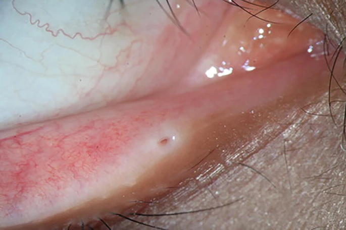
- Direct visual inspection of the puncta, comparing size and patency between the two eyes and against normal expected anatomy, often reveals visible narrowing, sometimes with the punctal opening reduced to a barely visible pinpoint or covered over by a thin membrane
- An increased tear meniscus height, reflecting reduced drainage
- A positive dye disappearance test (fluorescein instilled in the tear film clears slowly or incompletely) supporting a drainage problem, though this test alone does not localize the obstruction to the punctum specifically versus further downstream
- Difficulty or resistance passing a lacrimal probe or cannula through the punctum during attempted irrigation, which both confirms the stenosis and helps distinguish a punctal-level problem from an obstruction located further along the nasolacrimal duct
Differential Diagnosis
- Nasolacrimal duct obstruction (acquired, adult-onset) — obstruction further downstream, distinguished by irrigation testing showing the punctum itself is patent while fluid regurgitates or fails to pass further along the system
- Reflex hyperlacrimation from ocular surface irritation (dry eye, blepharitis, trichiasis) — the drainage system itself is entirely normal, and the problem is excess tear production rather than impaired drainage; distinguishing the two matters since the treatment approach is completely different
- Ectropion or punctal eversion — the punctum itself may be patent but positioned away from the tear lake, preventing normal tear drainage despite a structurally open drainage pathway, a mechanical positioning problem rather than true stenosis
- Floppy eyelid syndrome or other causes of lid malposition contributing to poor tear drainage mechanics, discussed in this site’s dedicated coverage of that condition
Evaluation
Careful external examination of the puncta, combined with lacrimal irrigation to assess patency and localize any obstruction along the drainage system, is usually sufficient to establish the diagnosis and distinguish punctal-level stenosis from more distal nasolacrimal duct obstruction.
It is also important to actively exclude reflex hyperlacrimation from ocular surface disease as a competing or coexisting explanation, because treating a mild degree of punctal narrowing surgically will not resolve tearing that is actually being driven by an underlying dry eye or blepharitis process, leaving the patient with the same symptom despite a technically successful procedure.
Management
Mild stenosis can sometimes be managed with punctal dilation alone, a simple in-office procedure using a graduated dilator to gently widen the punctal opening, though the effect can be temporary and repeat dilation is sometimes needed, making it a reasonable, low-risk first step before considering more definitive surgery.
More significant or recurrent stenosis is treated with a formal punctoplasty procedure, generally reserved for cases where simpler dilation has already proven inadequate.
Several surgical techniques exist, generally involving creating a small, controlled incision or excision to enlarge the punctal opening on a more durable basis than simple dilation provides.
Addressing any underlying contributing cause — switching a chronically used, poorly tolerated topical medication to a preservative-free alternative, or treating underlying chronic blepharitis — is a reasonable adjunct alongside mechanical treatment of the stenosis itself, because ongoing inflammation can otherwise contribute to recurrence even after a technically successful punctoplasty.
Patients should be counseled that surgical widening of the punctum, while generally effective, does not guarantee permanent resolution, and that some degree of recurrence over subsequent years is a recognized possibility that may eventually call for repeat treatment.


Document what you see
Two smartphone imaging tools built for everyday clinic use — one for the slit lamp, one for the fundus.
From Choroida — the team behind this siteReferences
- Kashkouli MB, Beigi B, Murthy R, Astbury N. Acquired external punctal stenosis: etiology and associated findings. American Journal of Ophthalmology.
- American Academy of Ophthalmology. Basic and Clinical Science Course, Section 7: Orbit, Eyelids, and Lacrimal System.
Test yourself
A few questions straight from this article.
-
What anatomical change defines punctal stenosis?
Punctal stenosis is narrowing or occlusion of the lacrimal punctum, the small opening at the medial eyelid margin through which tears drain into the nasolacrimal system. -
What is the most common cause of punctal stenosis in adults?
Age-related involutional change, with progressive fibrosis and narrowing of the punctal opening, is the most common cause of punctal stenosis. -
Which medication history should raise suspicion of punctal stenosis in a patient with new tearing?
Chronic use of certain glaucoma medications and other preserved topical drops is associated with punctal stenosis, an underrecognised side effect worth considering in long-term topical therapy. -
Which chemotherapy classes are recognised causes of punctal and canalicular stenosis?
Certain systemic chemotherapeutic agents, notably some taxane and fluoropyrimidine-based regimens, carry a recognised association with punctal and canalicular stenosis. -
Under which conditions do the symptoms of punctal stenosis characteristically worsen?
Wind, cold and outdoor conditions increase reflex tearing, which further overwhelms an already narrowed drainage pathway and makes symptoms worse. -
What is the main limitation of the dye disappearance test in suspected punctal stenosis?
A positive dye disappearance test, with fluorescein clearing slowly or incompletely, supports a drainage problem but cannot distinguish a punctal-level obstruction from one further downstream. -
What does lacrimal irrigation show in acquired nasolacrimal duct obstruction rather than punctal stenosis?
In nasolacrimal duct obstruction the punctum itself is patent while fluid regurgitates or fails to pass further along the system, whereas punctal stenosis causes resistance at the punctum itself. -
Which competing explanation must be excluded before operating on mild punctal narrowing?
Surgically treating mild punctal narrowing will not resolve tearing actually driven by dry eye or blepharitis, leaving the patient with the same symptom despite a technically successful procedure. -
What is the reasonable first step for mild punctal stenosis?
Mild stenosis can often be managed with punctal dilation, a simple low-risk in-office procedure, although the effect can be temporary and repeat dilation is sometimes needed before considering surgery. -
What should patients be told about the long-term outcome of punctoplasty for punctal stenosis?
Surgical widening of the punctum is generally effective but does not guarantee permanent resolution; some recurrence over subsequent years may eventually call for repeat treatment.
