
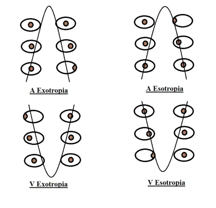
A-pattern and V-pattern strabismus describe a horizontal deviation that changes meaningfully in magnitude between upgaze and downgaze: converging (esotropia increasing, or exotropia decreasing) in upgaze and diverging in downgaze for A-pattern, and the opposite pattern for V-pattern.
Recognizing these patterns matters because they change the surgical plan beyond simply correcting the primary-position deviation.
Missing a significant pattern can leave a patient with persistent, function-limiting misalignment in reading or downgaze positions even after technically successful correction of the primary-position deviation.
These patterns are common enough in routine strabismus practice that a full motility assessment including upgaze and downgaze measurements should be considered a standard part of any comprehensive strabismus evaluation, not an optional addition reserved only for cases with an obvious craniofacial syndrome.
Defining the Patterns
- V-pattern — the horizontal deviation is more divergent (or less convergent) in upgaze than in downgaze, named for the V-shaped configuration this produces when the eye positions in up- and downgaze are plotted; a difference of at least 15 prism diopters between upgaze and downgaze is generally used as the threshold for clinical significance
- A-pattern — the opposite configuration, with the deviation more convergent (or less divergent) in upgaze than downgaze, using a threshold of at least 10 prism diopters difference between the two positions
Both patterns can occur with either esotropia or exotropia as the underlying primary-position deviation, giving four possible combinations (V-esotropia, V-exotropia, A-esotropia, A-exotropia), each with slightly different typical clinical associations and surgical considerations.
Underlying Mechanisms
The most widely accepted explanation involves oblique muscle dysfunction: overaction of the inferior oblique muscles is classically associated with V-pattern deviations, while overaction of the superior oblique muscles is classically associated with A-pattern deviations, reflecting how these muscles’ relative vertical and torsional actions shift the effective horizontal alignment differently in up- versus downgaze.
Other proposed contributing mechanisms include abnormal horizontal rectus muscle pulley positions and, in some craniofacial conditions, structurally abnormal orbital anatomy altering the effective vector of muscle pull.
Because more than one mechanism can plausibly contribute in a given patient, the surgical plan for a significant pattern is generally guided as much by the specific findings on careful motility examination — the degree of oblique overaction actually observed — as by any single presumed underlying cause.
All-fit Slit-Lamp Adapter
Record and share exactly what you see at the slit lamp. One adapter fits any slit lamp or surgical microscope — and any smartphone.
From Choroida — the team behind this siteAssociated Conditions
- Craniosynostosis syndromes (Crouzon, Apert, and related conditions) — V-pattern strabismus is particularly common in this group, related to the abnormal orbital and craniofacial bone structure
- Down syndrome — an association with A- and V-pattern strabismus has been reported
- Hydrocephalus and other conditions affecting the fourth ventricle or superior oblique function
- Idiopathic cases without an identified associated syndrome or structural cause, which represent a substantial proportion of cases seen in general pediatric ophthalmology practice
Clinical Evaluation
Measuring the horizontal deviation specifically in both upgaze (typically 25 degrees above primary position) and downgaze (25 degrees below), in addition to standard primary-position measurements, is essential to detect and quantify a pattern.
A pattern will simply be missed if only primary-position alignment is measured, which is why deliberately including these additional gaze positions in the strabismus exam matters.
Assessment for oblique muscle overaction (looking for the characteristic elevation of the adducting eye in inferior oblique overaction, or depression in superior oblique overaction) helps confirm the presumed underlying mechanism and directly informs surgical planning.
Because craniosynostosis and other associated syndromes carry their own distinct systemic implications, identifying a significant A- or V-pattern in a child with any suggestive craniofacial features should prompt broader evaluation for an associated syndrome rather than treating the strabismus as an isolated, unrelated finding.
Differential Diagnosis
- Simple horizontal strabismus without a significant pattern — the deviation remains relatively comitant across upgaze and downgaze, without meeting the diagnostic threshold for a true pattern
- Dissociated vertical deviation coexisting with horizontal strabismus — a distinct entity that can complicate the clinical picture and needs to be recognized and managed separately from a true A- or V-pattern
- Isolated oblique muscle palsy without an associated horizontal deviation pattern — vertical misalignment predominates rather than the gaze-dependent horizontal change that defines A- and V-pattern strabismus
Management
Surgical correction addresses both the primary-position horizontal deviation and the pattern itself, most commonly through vertical transposition of the horizontal rectus muscle insertions — moving the medial and/or lateral rectus insertions up or down relative to their standard position.
This shifts their relative effect between upgaze and downgaze and directly corrects the pattern.
When significant oblique muscle overaction is identified as the presumed mechanism, weakening the overacting oblique muscle (inferior oblique weakening for V-pattern, superior oblique weakening or a tenotomy-type procedure for A-pattern) is often combined with, or sometimes used instead of, transposition, depending on the specific findings and surgeon preference.
Because these patterns affect visual function in reading position (downgaze) and other everyday tasks involving up- or downgaze, surgical correction of a clinically significant, symptomatic pattern can meaningfully improve functional binocular vision even when the primary-position deviation alone was well tolerated or already addressed.
Postoperative follow-up specifically re-measures alignment in upgaze and downgaze, not just primary position, since a technically successful primary-position correction that leaves a significant residual pattern has not fully addressed the functional problem that originally prompted surgery, and any meaningful residual pattern is a reasonable indication for further surgical planning rather than being dismissed as an acceptable outcome.


Document what you see
Two smartphone imaging tools built for everyday clinic use — one for the slit lamp, one for the fundus.
From Choroida — the team behind this siteReferences
- von Noorden GK, Campos EC. Binocular Vision and Ocular Motility.
- Wright KW. Color Atlas of Strabismus Surgery: Strategies and Techniques.
- American Academy of Ophthalmology. Basic and Clinical Science Course, Section 6: Pediatric Ophthalmology and Strabismus.
Test yourself
A few questions straight from this article.
-
How is V-pattern strabismus defined in terms of horizontal deviation across vertical gaze?
In V-pattern the horizontal deviation is more divergent, or less convergent, in upgaze than in downgaze, producing the V-shaped configuration when eye positions are plotted. -
Which prism dioptre thresholds are generally used to call an A- or V-pattern clinically significant?
A difference of at least 15 prism dioptres between upgaze and downgaze is the usual threshold for a clinically significant V-pattern, while at least 10 prism dioptres is used for A-pattern. -
Overaction of which muscles is classically associated with V-pattern strabismus?
Inferior oblique overaction is classically associated with V-pattern deviations, while superior oblique overaction is classically associated with A-pattern deviations. -
Which examination finding indicates inferior oblique overaction in a patient with pattern strabismus?
Inferior oblique overaction produces characteristic elevation of the adducting eye, whereas superior oblique overaction produces depression; identifying this confirms the presumed mechanism and informs surgical planning. -
At which gaze positions should the horizontal deviation be measured to detect A- and V-pattern strabismus?
Measuring the horizontal deviation in upgaze and downgaze, typically 25 degrees above and below primary position, is essential; a pattern is simply missed if only primary-position alignment is measured. -
In which group of conditions is V-pattern strabismus particularly common?
V-pattern strabismus is particularly common in craniosynostosis syndromes including Crouzon and Apert, related to the abnormal orbital and craniofacial bone structure. -
Which surgical manoeuvre most commonly corrects the pattern itself in A- and V-pattern strabismus?
Moving the medial and/or lateral rectus insertions up or down relative to their standard position shifts their relative effect between upgaze and downgaze, directly correcting the pattern. -
Which oblique muscle procedure is used when significant oblique overaction underlies an A-pattern?
When superior oblique overaction is the presumed mechanism of an A-pattern, superior oblique weakening or a tenotomy-type procedure is combined with, or sometimes replaces, transposition surgery. -
What distinguishes simple horizontal strabismus from true A- or V-pattern strabismus?
In simple horizontal strabismus the deviation remains relatively comitant between upgaze and downgaze and never meets the diagnostic threshold required to call it a true pattern. -
What must postoperative follow-up after pattern strabismus surgery specifically include?
A primary-position correction that leaves a significant residual pattern has not addressed the functional problem, so follow-up re-measures upgaze and downgaze; a meaningful residual pattern justifies further surgical planning.
