
A cataract operation is expected to last a lifetime, and for most people it does.
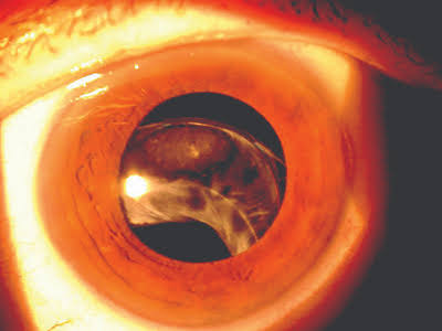
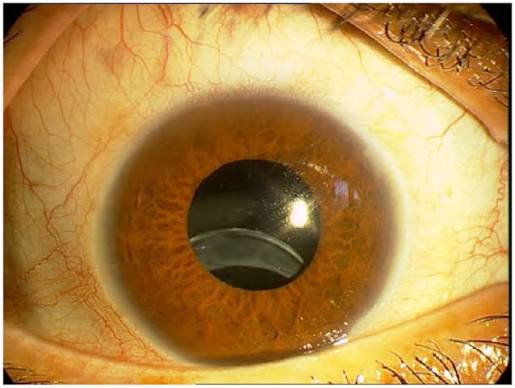
In a small proportion of eyes, though, the capsular bag containing the lens implant slowly drifts out of position, years or even decades after surgery, because the zonules supporting it have failed.
This is late in-the-bag intraocular lens dislocation, and it has become the leading cause of late IOL dislocation in many reports.
What actually happens
In late in-the-bag dislocation, the IOL stays inside the capsular bag, but the whole bag, along with its zonular attachments, comes loose.
The bag may sublux inferiorly, or it may fall into the vitreous.
The lens optic is displaced, causing blur or double vision.
This is different from early dislocation, caused by an intraoperative problem, where the IOL is placed outside the bag or the haptic slips out.
The zonules degrade slowly with time in conditions that weaken them, and the contraction of the capsular bag (phimosis) adds a stress.
Who is at risk
- Pseudoexfoliation, by far the commonest association (see pseudoexfoliation syndrome)
- Prior trauma
- High axial myopia and previous vitreoretinal surgery
- Retinitis pigmentosa
- Uveitis
- Connective tissue disorders such as Marfan syndrome
- Long time since surgery. Population-based work found a cumulative incidence of roughly 0.1 percent at 5 years, rising to about 1.7 percent at 25 years
Surgeons who recognize weak zonules at the time of the original surgery can place a capsular tension ring, which may reduce the risk (see capsular tension ring indications and zonular dehiscence in cataract surgery).
All-fit Slit-Lamp Adapter
Record and share exactly what you see at the slit lamp. One adapter fits any slit lamp or surgical microscope — and any smartphone.
From Choroida — the team behind this siteHow it presents
- Gradual or sudden blur, often with a change in refraction
- Monocular diplopia, or a visible edge of the lens in the pupil
- Glare, if the lens edge crosses the visual axis
- Positional changes in vision when lying down, if the lens falls back
- Pain and redness if raised pressure or uveitis develop
Examination at the slit lamp, with a dilated pupil, will show the bag and lens displaced, with a clear zone where the bag used to be.
Phacodonesis and iridodonesis are common.
The lens may be tilted, decentered, or fully in the vitreous.
Management options
The right approach depends on the position of the bag, the patient’s vision, and the state of the retina.
- Observation if the lens is slightly decentered and vision is good
- Repositioning and suturing the bag or haptics to the sclera or iris
- IOL exchange, where the dislocated lens is removed and a new lens is placed with scleral or iris fixation, or an anterior chamber lens is used when support is limited (see scleral-fixated intraocular lens)
- Pars plana vitrectomy if the lens is in the vitreous, with attention to retinal tears
Whatever the approach, the eye should be assessed for retinal tears, cystoid macular edema, and pressure elevation.
Complications
Retinal detachment, vitreous hemorrhage, cystoid macular edema, glaucoma, suture exposure or breakage with subsequent re-dislocation, and endophthalmitis are known complications.
Patients with pseudoexfoliation need long-term follow-up since the underlying disease continues to progress.
What the patient should be told
Patients with pseudoexfoliation, trauma, or high myopia who have had cataract surgery should know that a late change in vision, double vision, or a visible edge of the lens in the pupil needs prompt review. They should avoid heavy blows to the head and be aware of the symptoms. Explain that the problem is the support of the lens and not the lens implant itself, and that repair is possible.
Surgical planning
The surgeon needs to decide whether to reposition the bag with its lens, to remove the bag and lens and replace it, or to leave the lens and observe. Factors include the position of the lens, the state of the zonules, the condition of the cornea, the retina, and the glaucoma status. A careful vitrectomy is needed when the lens is in the vitreous, and the peripheral retina should be examined for tears. The choice of fixation method, with scleral or iris suturing or with glued haptics, depends on the surgeon's experience and the anatomy.
Follow-up
After surgery, review for pressure rises, macular edema, retinal tears, and suture problems. Long-term follow-up is advisable because the underlying disease, such as pseudoexfoliation, continues to progress.
Prognosis
With appropriate surgery, visual results are good in most cases.
The best defense is early recognition of zonular weakness before the first operation.


Document what you see
Two smartphone imaging tools built for everyday clinic use — one for the slit lamp, one for the fundus.
From Choroida — the team behind this siteReferences
- Pueringer SL, Hodge DO, Erie JC. Risk of late intraocular lens dislocation after cataract surgery, 1980-2009: a population-based study. Am J Ophthalmol. 2011;152:618-623.
- Gimbel HV, Condon GP, Kohnen T, Olson RJ, Halkiadakis I. Late in-the-bag intraocular lens dislocation: incidence, prevention, and management. J Cataract Refract Surg. 2005;31:2193-2204.
- Davis D, Brubaker J, Espandar L, et al. Late in-the-bag spontaneous intraocular lens dislocation: evaluation of 86 consecutive cases. Ophthalmology. 2009;116:664-670.
- Hayashi K, Hirata A, Hayashi H. Possible predisposing factors for in-the-bag and out-of-the-bag intraocular lens dislocation and outcomes of intraocular lens exchange surgery. Ophthalmology. 2007;114:969-975.
