
Iris prolapse is the protrusion of iris tissue through a hole in the cornea or at the limbus.
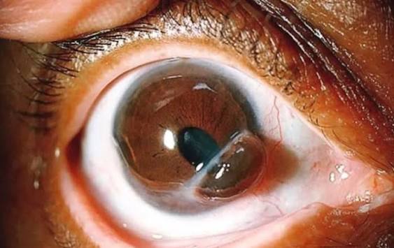
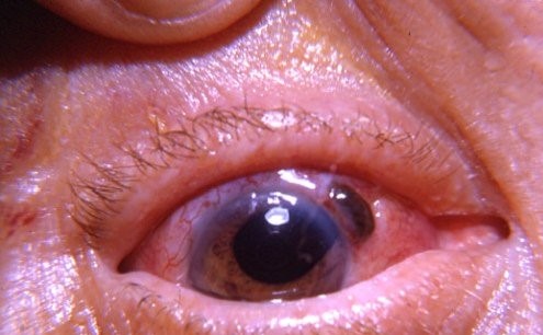
The iris is drawn into the wound by the outflow of aqueous, and it plugs it.
It is seen after open-globe injuries, as a complication of surgery, and in some eyes with a weak or thinned cornea.
It needs prompt attention, since the eye is open, and there is a danger of infection.
What it looks like
- A dark brown or black knuckle of tissue at the wound, which may be covered with fibrin or blood
- A pupil that is peaked or pulled toward the wound, often teardrop-shaped
- A shallow or flat anterior chamber
- A leaking wound that is Seidel positive, although the iris can plug the leak
- Pain, photophobia, and reduced vision
Prolapsed iris must be distinguished from a pigmented foreign body, a conjunctival nevus, or a staphyloma.
Common causes
- Penetrating and perforating injuries, with a corneal or corneoscleral laceration
- Surgical wounds after cataract surgery, with unstable or leaking incisions, particularly in eyes with intraoperative floppy iris syndrome (see floppy iris syndrome)
- Corneal ulcer perforation in infectious keratitis or melts (see descemetocele)
- Wound dehiscence after keratoplasty or other surgery (see corneal wound dehiscence)
- Raised pressure from a Valsalva maneuver or a blow to the eye in the early postoperative period
All-fit Slit-Lamp Adapter
Record and share exactly what you see at the slit lamp. One adapter fits any slit lamp or surgical microscope — and any smartphone.
From Choroida — the team behind this siteWhy it is dangerous
The open wound provides a path for microbes, so the risk of endophthalmitis is high.
The prolapsed iris may become ischemic and necrotic.
Chronic prolapse can lead to epithelial downgrowth, to anterior synechiae and glaucoma, and, after penetrating injury, there is a rare risk of sympathetic ophthalmia (see sympathetic ophthalmia).
Initial management
- Protect the eye with a rigid shield, and do not press on it
- Avoid manipulation of the globe, and give systemic antibiotics and tetanus prophylaxis as needed
- Keep the patient fasting for surgery, and control pain and vomiting
- Obtain imaging to exclude an intraocular foreign body, with CT, if there is a history of injury
- Arrange urgent surgical repair
Surgical principles
- Recent prolapse (within about 24 to 48 hours), clean and viable: the iris can be reposited into the anterior chamber with a viscoelastic and a spatula, after the wound is irrigated, and the wound is sutured.
- Old prolapse, necrotic, contaminated, or adherent: excise the exposed iris, carefully avoiding traction on the iris root, and close the wound.
- Suture the wound with nylon, in watertight closure, and form the chamber.
- Give intracameral or intravitreal antibiotics when there is a high risk of infection.
Surgery for cataract wounds is similar, and may need wound reinforcement or a repeat of the incision.
Follow-up
- Watch for endophthalmitis in the first week
- Treat inflammation with topical steroids and cycloplegics
- Check for glaucoma, cataract, and retinal problems
- Plan any pupil or iris reconstruction after healing
Examination and classification
Examine the wound with the slit lamp, using fluorescein to check for leakage, and note the extent of the wound, the tissue prolapsed, and the depth of the anterior chamber. Look for lens damage, hyphema, and signs of infection. If the globe is obviously open, avoid pressure, protect the eye with a shield, and do not attempt to push the iris back at the bedside. Obtain a CT scan in trauma to look for a foreign body, and note the time of the injury and the tetanus status.
The decision to reposit or excise
Fresh, clean, viable iris that has been outside the eye for a short time can be reposited, with a viscoelastic and a spatula through a separate paracentesis. Iris that is dark, dull, or has been exposed for more than a day or two is likely to be contaminated and devitalized, and it should be excised. The surgeon should aim to preserve as much healthy iris as possible, and avoid tearing the iris root.
Antibiotic cover and follow-up
Systemic and sometimes intracameral antibiotics are used, and the patient should be examined daily in the first week. Watch for endophthalmitis, uveitis, raised pressure, and hyphema, and plan for secondary procedures such as pupilloplasty or cataract surgery once the eye is quiet.
Prevention
Careful wound construction in cataract surgery, with a long enough tunnel and a watertight closure, reduces the risk. In patients with floppy iris syndrome, pupil expanders and modified techniques help, and intracameral medications reduce the problem. Protective eyewear prevents many traumatic injuries, and patients should be advised to avoid heavy straining and rubbing in the early postoperative period.
Prognosis
With early repair, outcomes are good in cases of clean wounds.
Delayed treatment, a contaminated wound, or associated posterior segment injury worsens the prognosis.


Document what you see
Two smartphone imaging tools built for everyday clinic use — one for the slit lamp, one for the fundus.
From Choroida — the team behind this siteReferences
- Kuhn F, ed. Ocular Traumatology. Berlin: Springer; 2008.
- Chang DF, Campbell JR. Intraoperative floppy iris syndrome associated with tamsulosin. J Cataract Refract Surg. 2005;31:664-673.
- Kanski JJ, Bowling B. Clinical Ophthalmology: A Systematic Approach. 7th ed. London: Elsevier; 2011.
- Yanoff M, Duker JS. Ophthalmology. 5th ed. Philadelphia: Elsevier; 2018.
