

Unilateral Pigmentary Retinopathy DISEASE entity
Unilateral Pigmentary Retinopathy (UPR), sometimes also referred to as unilateral retinitis pigmentosa) refers to a rare, sporadic disorder that involves degeneration and atrophy of the retina in one eye, specifically at the level of the photoreceptors.
It can mimic the appearance of Retinitis Pigmentosa (RP); however, Unilateral Pigmentary Retinopathy (UPR) typically presents in one eye with a normal fellow eye whereas RP presents bilaterally.
Prevalence is approximately 1 in 4000 with less than 100 cases reported in the literature.
The exact etiology of Unilateral Pigmentary Retinopathy (UPR) is unknown. It is thought that somatic mutation during embryogenesis involves cells forming the retina and RPE (retinal pigment epithelium).
Due to the mutation occurring early in embryogenesis, it has the potential to also affect germ cell lines and thus poses minimal risk of passing on the mutation to subsequent offspring.
Unilateral Pigmentary Retinopathy (UPR) can be inherited in an autosomal dominant, autosomal recessive, or X-linked recessive pattern. Cases have been reported with p.R677X germline mutation in the RP1 gene, USH2AW4149R mutation, and PDE6B mutation.
The nature of the pigmentary changes may not reliably predict the diagnosis or functional phenotype.
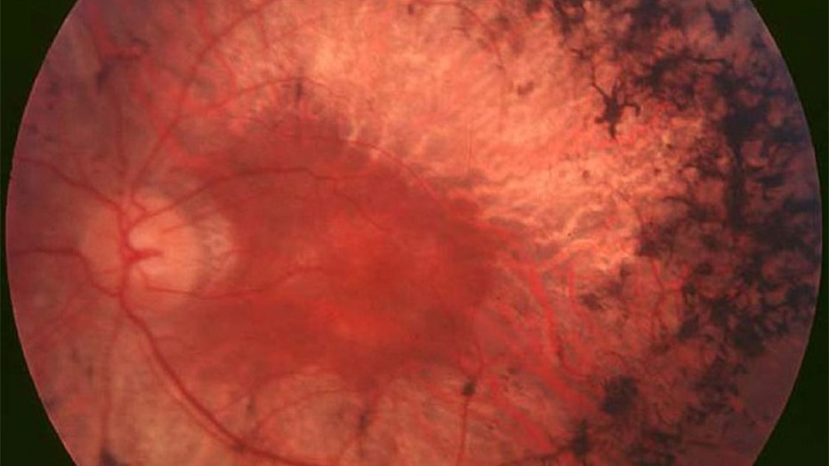
Intraretinal pigmentary deposition, RPE depigmentation, disc pallor, and vessel attenuation can occur in Unilateral Pigmentary Retinopathy (UPR) depending on the stage of the disease but are not pathognomonic.
Mechanisms for the fundal appearance seen in RP of bone spicules have been studied in mouse models. The pathophysiology of bone spicule formation seen in UPR occurs due to photoreceptor cell death by apoptosis.
This eventually leads to the degeneration of the outer retina, causing direct contact with the inner retinal vessels and migration of the RPE cells.
Extracellular matrix deposits form as a result of the mislocalized RPE cells that partially seal the contacting vessels.
Physical examination
Visual acuity: Patients present with decreased visual acuity at night (nyclopia); however, visual acuity can be variable, as cases have been reported where visual acuity has ranged from 20/15 to counting fingers.
Fundus exam may reveal mottling of the retinal pigment epithelium (RPE), bone-spicule formations, granular pigmentary disturbances or punched-out atrophic RPE lesions, attenuated retinal vessels, and waxy pallor of the optic disc (Figure 1).
Visual field examination: Peripheral visual field loss is common.
Fundus Explorer Pro
Photograph the retinal findings described here with the phone already in your pocket — 22 D optics and built-in illumination in one handheld unit.
From Choroida — the team behind this siteUnilateral Pigmentary Retinopathy MANAGEMENT
General treatment
There is no gold standard or definite treatment for patients with Unilateral Pigmentary Retinopathy (UPR). Supportive treatment is the standard of care. Commonly utilized treatments have included antioxidants, vitamins, and nutritional supplements but have not shown true benefit.

Associated conditions, such as cystoid macular edema (CME), should be managed with appropriate medical therapy – typically, carbonic anhydrase inhibitors.
However, CME due to intraocular inflammation does not improve with this treatment which might respond to intravitreal, periocular, or oral steroids.
Medical follow up
Medical follow-up should be done to monitor the progression of the disease in the affected eye as well as monitor the fellow eye to ensure the patient is not presenting with an asymmetric form of RP.
Annual ocular examinations are recommended to monitor visual acuity and Goldmann’s visual field.


Document what you see
Two smartphone imaging tools built for everyday clinic use — one for the slit lamp, one for the fundus.
From Choroida — the team behind this siteREFERENCES
- Weiss, J.F. and R.J. Nicholl, Nonsyphilitic unilateral retinitis pigmentosa. Am J Ophthalmol, 1968. 65(4): p. 573-4.
- Carr, R.E. and I.M. Siegel, Unilateral retinitis pigmentosa. Arch Ophthalmol, 1973. 90(1): p. 21-6.
- Chen, H., et al., Unilateral retinitis pigmentosa with amblyopia in the fellow eye. Graefes Arch Clin Exp Ophthalmol, 2006. 244(12): p. 1701-4.
- Grisanti, S., et al., Unilateral pigmentary degeneration of the retina associated with heterochromia iridis.Graefes Arch Clin Exp Ophthalmol, 1998. 236(12): p. 940-4.
- Spadea, L., et al., Unilateral retinitis pigmentosa: clinical and electrophysiological report of four cases.Ophthalmologica, 1998. 212(5): p. 350-4.
- Berson, E.L., Retinitis pigmentosa. The Friedenwald Lecture. Invest Ophthalmol Vis Sci, 1993. 34(5): p. 1659-76.
Test yourself
A few questions straight from this article.
-
In unilateral pigmentary retinopathy, retinal degeneration and atrophy occur at which level?
Unilateral pigmentary retinopathy is a rare sporadic disorder in which degeneration and atrophy of the retina occur specifically at the photoreceptor level. -
Which feature separates unilateral pigmentary retinopathy from classic retinitis pigmentosa?
Although it mimics retinitis pigmentosa, unilateral pigmentary retinopathy typically affects one eye with a normal fellow eye, whereas retinitis pigmentosa presents bilaterally. -
Which mechanism is thought to underlie unilateral pigmentary retinopathy?
The exact etiology is unknown, but a somatic mutation arising during embryogenesis in the cells forming the retina and retinal pigment epithelium is the proposed mechanism. -
Bone-spicule formation in unilateral pigmentary retinopathy is initiated by which event?
Apoptotic photoreceptor death leads to outer retinal degeneration, which brings the retina into direct contact with inner retinal vessels and drives migration of RPE cells. -
Across reported cases of unilateral pigmentary retinopathy, visual acuity has spanned which range?
Visual acuity in unilateral pigmentary retinopathy is highly variable, with reported cases ranging from 20/15 down to counting fingers. -
Which fundus picture is described in unilateral pigmentary retinopathy?
Fundus examination may show RPE mottling, bone-spicule formation, granular pigmentary disturbance or punched-out atrophic RPE lesions, attenuated retinal vessels and waxy optic disc pallor. -
Which visual field abnormality is common in unilateral pigmentary retinopathy?
Peripheral visual field loss is the common finding on visual field examination in unilateral pigmentary retinopathy. -
What constitutes the standard of care in unilateral pigmentary retinopathy?
There is no gold standard or definite treatment for unilateral pigmentary retinopathy; supportive care is standard, and antioxidants, vitamins and nutritional supplements have not shown true benefit. -
Cystoid macular edema complicating unilateral pigmentary retinopathy is typically managed with which agents?
Cystoid macular edema associated with unilateral pigmentary retinopathy is typically treated with carbonic anhydrase inhibitors; edema driven by intraocular inflammation does not respond and may require steroids. -
Why is the unaffected fellow eye followed in unilateral pigmentary retinopathy?
Follow-up monitors progression in the affected eye and watches the fellow eye to confirm the patient does not in fact have an asymmetric form of retinitis pigmentosa; annual exams track acuity and Goldmann visual fields.

Writes and reviews the clinical articles on Educate. Every post is checked against peer-reviewed sources before it goes live.


