
A patient who had LASIK the day before returns with a hazy, gritty-looking cornea.
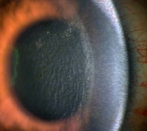
The surface looks fine, but under the flap there is a fine, granular, white haze, like windblown sand.
That is diffuse lamellar keratitis, or DLK, popularly called “sands of the Sahara.” It is sterile, and it responds well to treatment when it is caught early.
What causes it
DLK is an inflammatory response in the flap interface triggered by material that should not be there.
Reported triggers include bacterial endotoxins from sterilization equipment, meibomian gland secretions, debris from the microkeratome or the laser, talc from surgical gloves, preservatives, and povidone-iodine.
In many outbreaks, a contaminated cleaning solution or sterilizer was found.
Inflammatory cells enter the interface through the limbal vessels and the tear film, and the result is a diffuse inflammatory reaction.
Presentation and timing
- Typically appears in the first 1 to 3 days after surgery, with a peak at day 2 to 3
- Mild cases have few symptoms. Others report blur, haze, photophobia, or mild discomfort
- Pain, if it is marked, should raise suspicion of infection or another cause
- At the slit lamp there are white, granular cells in the interface, starting at the periphery and often more evident near the flap edge
All-fit Slit-Lamp Adapter
Record and share exactly what you see at the slit lamp. One adapter fits any slit lamp or surgical microscope — and any smartphone.
From Choroida — the team behind this siteFour stages
A commonly used grading scheme (Linebarger) divides DLK as follows:
- Stage 1: white, granular cells in the periphery of the flap, not involving the visual axis
- Stage 2: cells extend into the central cornea
- Stage 3: denser aggregation of cells centrally, with relative clearing of the periphery, and haze that begins to reduce vision
- Stage 4: stromal melting with scarring, and possible permanent loss of vision
The aim of treatment is to prevent progression from stages 1 and 2 to stage 3 or 4.
Differential diagnosis
- Infectious keratitis, including atypical mycobacteria and fungi, develops later, with focal infiltrates, pain, and redness, and needs urgent culture and antimicrobial therapy
- Epithelial ingrowth, which develops later and is nodular (see epithelial ingrowth after LASIK)
- Pressure-induced stromal keratitis, in patients on steroids with high intraocular pressure, where fluid accumulates in the interface
- Interface debris, which is non-inflammatory and does not progress
Treatment
- Stage 1: frequent topical steroids, for example hourly while awake, with close follow-up
- Stage 2: the same, with a short course of oral steroids if progression occurs
- Stage 3 and 4: lift the flap and irrigate the interface with saline to remove inflammatory cells and debris, followed by intensive steroids
Tapering must be gradual.
Monitor intraocular pressure carefully, since steroids can raise it, and a high pressure can itself cause interface fluid.
Prevention
- Strict protocols for cleaning and sterilizing instruments, with fresh water and detergents with no endotoxin
- Care with gloves and drapes
- Meibomian gland and lid margin management before surgery
- Reporting clusters of cases early, since they usually point to a common source
How the first-day visit should go
Examine every post-LASIK patient on the first day with the slit lamp, looking at the flap interface for white granular cells, particularly at the periphery. Note the grade, record the visual acuity and the pressure, and give a clear plan. Early recognition of stage 1 allows treatment with frequent steroids before the process reaches the center of the cornea. If a cluster of cases appears on the same day or from the same operating room, report it immediately to the surgical team so that the source can be investigated.
Treatment details
Topical corticosteroids such as prednisolone acetate are used frequently, at intervals of one to two hours during the day in the early stages, with a gradual taper over one to two weeks. In more severe cases, oral steroids and interface irrigation are added. Irrigation lifts the flap and washes out the inflammatory cells, and it should be performed early in stage 3 before melting begins. Steroid response with raised pressure must be monitored, because it can cause interface fluid and confuse the picture.
How to explain it
Tell the patient that the haze is an inflammatory reaction to something that got under the flap, not an infection, that it usually responds to drops, and that frequent follow-up in the first days is a crucial part of the treatment.
Special situations
In patients with dry eye or blepharitis, treat the lid margin before surgery. After an enhancement, a repeat check for DLK on the first day is advised, since the interface is again exposed.
Prognosis
Most cases resolve fully without sequelae when treated early.
Delayed or advanced cases, especially stage 4, can leave scars and irregular astigmatism.


Document what you see
Two smartphone imaging tools built for everyday clinic use — one for the slit lamp, one for the fundus.
From Choroida — the team behind this siteReferences
- Smith RJ, Maloney RK. Diffuse lamellar keratitis. A new syndrome in lamellar refractive surgery. Ophthalmology. 1998;105:1721-1726.
- Linebarger EJ, Hardten DR, Lindstrom RL. Diffuse lamellar keratitis: diagnosis and management. J Cataract Refract Surg. 2000;26:1072-1077.
- Johnson JD, Harissi-Dagher M, Pineda R, Yoo S, Azar DT. Diffuse lamellar keratitis: incidence, associations, outcomes, and a new classification system. J Cataract Refract Surg. 2001;27:1560-1566.
- Ambrosio R Jr, Wilson S. Complications of laser in situ keratomileusis: etiology, prevention, and treatment. J Refract Surg. 2001;17:350-379.
Test yourself
A few questions straight from this article.
-
Diffuse lamellar keratitis after LASIK is best described as which process?
DLK is a sterile inflammatory response in the flap interface triggered by material that should not be there, producing the fine granular haze nicknamed sands of the Sahara. -
Which trigger has been implicated in outbreaks of diffuse lamellar keratitis?
Reported triggers include bacterial endotoxins from sterilization equipment, meibomian secretions, microkeratome or laser debris, glove talc, preservatives and povidone-iodine, and many outbreaks were traced to a contaminated cleaning solution or sterilizer. -
When does diffuse lamellar keratitis typically appear after LASIK?
DLK typically appears in the first 1 to 3 days after surgery with a peak at day 2 to 3, which is why every post-LASIK patient is examined on the first day. -
What should marked pain in a patient with suspected diffuse lamellar keratitis raise?
DLK causes at most mild discomfort, blur, haze or photophobia, so marked pain should raise suspicion of infection or another cause. -
What defines stage 1 diffuse lamellar keratitis in the Linebarger scheme?
Stage 1 is white granular cells in the periphery of the flap without involvement of the visual axis, and recognising it allows treatment before the process reaches the centre. -
What characterizes stage 4 diffuse lamellar keratitis?
Stage 4 is stromal melting with scarring and possible permanent loss of vision, which is why treatment aims to stop progression from stages 1 and 2. -
Which feature favours infectious keratitis over diffuse lamellar keratitis after LASIK?
Infectious keratitis, including atypical mycobacteria and fungi, develops later with focal infiltrates, pain and redness, and demands urgent culture and antimicrobial therapy. -
What is the treatment of stage 1 diffuse lamellar keratitis?
Stage 1 is treated with frequent topical steroids, for example hourly while awake, with close follow-up and a gradual taper. -
What is added in the treatment of stage 3 or stage 4 diffuse lamellar keratitis?
In stages 3 and 4 the flap is lifted and the interface irrigated with saline to wash out inflammatory cells and debris, followed by intensive steroids; irrigation should be done early in stage 3, before melting begins. -
Why must intraocular pressure be watched during steroid treatment of diffuse lamellar keratitis?
Steroids can raise intraocular pressure, and a high pressure can itself produce interface fluid that confuses the clinical picture.

Writes and reviews the clinical articles on Educate. Every post is checked against peer-reviewed sources before it goes live.


