
Chronic postoperative endophthalmitis is a delayed, indolent form of intraocular infection following cataract or other intraocular surgery, presenting weeks to months after the original procedure rather than in the acute postoperative window discussed in relation to acute postoperative endophthalmitis in its own dedicated article on this site.
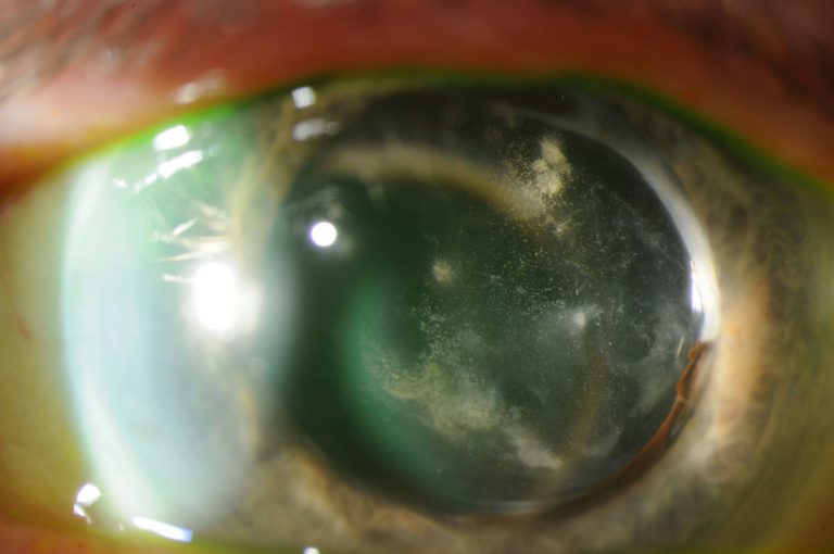
Its slow, smoldering course and often subtle presentation make it a genuinely different clinical problem from acute endophthalmitis, frequently mistaken for chronic postoperative inflammation or recurrent uveitis before the correct infectious diagnosis is finally reached.
Recognizing this distinct pattern matters because the causative organisms, workup, and treatment approach for chronic endophthalmitis differ meaningfully from those used for the far more dramatic and rapidly progressive acute form.
Chronic endophthalmitis is genuinely uncommon compared to acute postoperative infection, but its slow, easily misattributed course means the true time from surgery to correct diagnosis is often considerably longer than for the acute form.
Why It Looks So Different From Acute Endophthalmitis
Chronic postoperative endophthalmitis is most classically caused by Cutibacterium acnes, formerly known as Propionibacterium acnes, a low-virulence organism capable of surviving sequestered within the capsular bag for extended periods without provoking the severe, rapid inflammatory response typical of more virulent acute pathogens.
This sequestration within the capsular bag, often behind the intraocular lens where it is relatively protected from both the immune system and topical antibiotic penetration, is what allows the infection to persist smolderingly for weeks to months rather than declaring itself acutely.
Fungal organisms can also cause a similarly indolent, chronic presentation, and should be considered alongside Cutibacterium acnes in the differential for any low-grade, persistent postoperative inflammation that does not fit a typical acute infectious or purely inflammatory pattern.
Clinical Presentation
- Chronic, low-grade anterior chamber inflammation developing weeks to months after otherwise uneventful cataract surgery, often initially responsive to topical corticosteroids but recurring when they are tapered
- A characteristic white plaque or granulomatous deposit on the posterior capsule, when present, a relatively specific finding for Cutibacterium acnes infection
- Mild to moderate vision loss and discomfort, generally far less dramatic than the pain and rapid vision loss typical of acute endophthalmitis
- A pattern of repeated response to steroid treatment followed by recurrence upon tapering, which is one of the more important clues distinguishing this from simple postoperative inflammation without an infectious component
All-fit Slit-Lamp Adapter
Record and share exactly what you see at the slit lamp. One adapter fits any slit lamp or surgical microscope — and any smartphone.
From Choroida — the team behind this siteWhy It Is Often Misdiagnosed
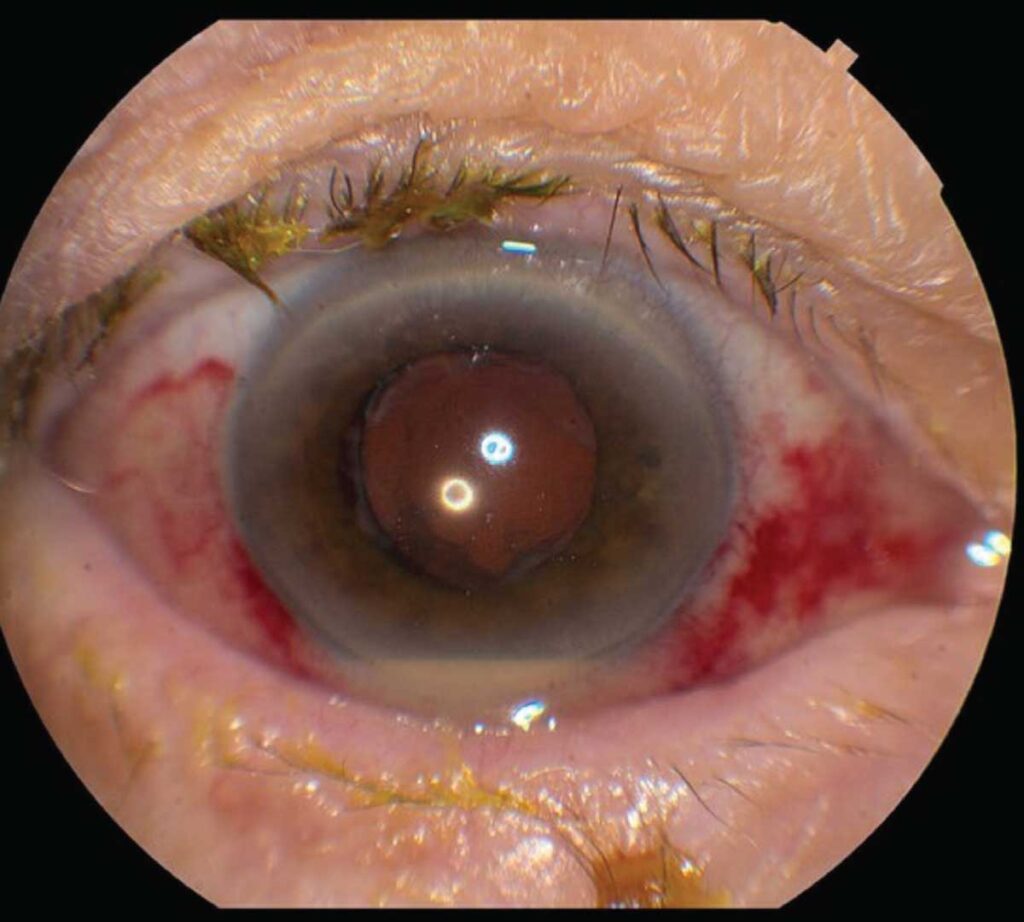
The relatively mild, smoldering presentation, without the severe pain, marked vision loss, and hypopyon typical of acute endophthalmitis, means chronic endophthalmitis often does not trigger the same immediate suspicion for infection.
Repeated response to corticosteroid treatment, followed by recurrence each time steroids are tapered, is frequently and understandably interpreted initially as simple postoperative or recurrent idiopathic uveitis rather than an underlying, persistent low-grade infection.
This recurring steroid-response pattern, rather than any single dramatic finding, is often the clue that should eventually prompt consideration of chronic endophthalmitis, particularly when the pattern repeats over multiple treatment cycles without ever fully resolving.
Diagnostic Evaluation
Aqueous or vitreous sampling for culture is needed to confirm the diagnosis, though a negative culture does not fully exclude chronic endophthalmitis given the fastidious, slow-growing nature of Cutibacterium acnes, which sometimes requires extended culture incubation to grow.
Polymerase chain reaction testing of intraocular fluid can improve detection sensitivity compared to culture alone, particularly useful given how often standard cultures fail to grow this specific organism even when it is truly present.
A high index of clinical suspicion, based on the characteristic recurring pattern described above, often drives the decision to proceed with treatment even when microbiologic confirmation remains elusive or delayed.
Management
Partial treatment with intravitreal antibiotics alone frequently fails to achieve lasting cure, since organisms sequestered within the capsular bag are often incompletely reached by antibiotics circulating in the vitreous cavity.
Definitive treatment typically requires removal of the capsular bag along with the intraocular lens, or at minimum a partial capsulectomy, to physically eliminate the sequestered focus of infection rather than relying on antibiotics alone to eradicate it.
Intravitreal antibiotics are used alongside surgical intervention, and vitrectomy is often performed at the same time as capsular bag and lens removal to address any associated vitreous involvement comprehensively.
Visual prognosis after appropriately targeted treatment is generally favorable given the organism’s low virulence, in clear contrast to the often guarded prognosis associated with acute endophthalmitis from more aggressive pathogens.


Document what you see
Two smartphone imaging tools built for everyday clinic use — one for the slit lamp, one for the fundus.
From Choroida — the team behind this siteReferences
- Clark WL, Kaiser PK, Flynn HW Jr, et al. Treatment strategies and visual acuity outcomes in chronic postoperative Propionibacterium acnes endophthalmitis. Ophthalmology.
- American Academy of Ophthalmology. Basic and Clinical Science Course, Section 12: Retina and Vitreous.
- Aldave AJ, Stein JD, Deramo VA, et al. Treatment strategies for postoperative Propionibacterium acnes endophthalmitis. Ophthalmology.
Test yourself
A few questions straight from this article.
-
When does chronic postoperative endophthalmitis typically declare itself after intraocular surgery?
Chronic postoperative endophthalmitis is a delayed, indolent infection appearing weeks to months after cataract or other intraocular surgery, well outside the acute postoperative window. -
Which organism most classically causes chronic postoperative endophthalmitis?
Cutibacterium acnes, formerly called Propionibacterium acnes, is the classic cause. Its low virulence lets it persist without provoking the severe rapid inflammation of more aggressive pathogens. -
Where does the causative organism of chronic postoperative endophthalmitis characteristically sequester itself?
Sequestration inside the capsular bag, typically behind the intraocular lens, shelters the organism from both the immune system and topical antibiotic penetration, allowing a smouldering course. -
Which examination finding is relatively specific for Cutibacterium acnes endophthalmitis?
When present, a white plaque or granulomatous deposit on the posterior capsule is a relatively specific sign of Cutibacterium acnes infection in this setting. -
Which pattern should raise suspicion of chronic endophthalmitis rather than simple postoperative inflammation?
A cycle of responding to corticosteroids and then relapsing each time they are tapered, repeating over several treatment courses without ever fully resolving, is the key clue. -
Why may intraocular fluid culture be negative despite genuine chronic endophthalmitis?
Cutibacterium acnes is fastidious and slow-growing, sometimes needing extended incubation, so a negative culture does not exclude chronic postoperative endophthalmitis. -
What advantage does polymerase chain reaction testing of intraocular fluid offer in chronic endophthalmitis?
Polymerase chain reaction testing of intraocular fluid raises detection sensitivity above culture alone, which matters because standard cultures often fail to grow this organism. -
Why do intravitreal antibiotics alone frequently fail to cure chronic postoperative endophthalmitis?
Antibiotic circulating in the vitreous cavity often fails to reach organisms sequestered within the capsular bag, so partial treatment with injections alone rarely achieves lasting cure. -
What does definitive treatment of chronic postoperative endophthalmitis typically require?
Definitive treatment physically eliminates the sequestered focus by removing the capsular bag with the intraocular lens, or at minimum performing a partial capsulectomy, with intravitreal antibiotics alongside. -
What is the visual prognosis after appropriately targeted treatment of chronic postoperative endophthalmitis?
Because the causative organism has low virulence, visual prognosis after targeted treatment is generally favourable, contrasting with the often guarded outlook in acute endophthalmitis.
