
A conjunctival nevus is a benign melanocytic lesion of the conjunctiva, and it is by far the most common pigmented conjunctival lesion encountered in clinic.
Most are picked up incidentally, either by the patient noticing a spot on the eye or during a routine examination for something unrelated, and the great majority never need anything more than reassurance and periodic monitoring.
The clinical task is a familiar one in ophthalmology: separating the common, harmless lesion from the rare one that needs closer attention, in this case conjunctival melanoma or primary acquired melanosis with atypia.
What Is a Conjunctival Nevus?
Conjunctival nevi are congenital or acquired proliferations of nevus cells within the conjunctival epithelium and substantia propria.
Most become clinically apparent in the first two decades of life, often becoming more noticeably pigmented around puberty as hormonal changes affect melanocyte activity, which can understandably worry a patient or parent who perceives this as new growth rather than simply increased pigmentation of an existing lesion.
Nevi are classified histologically by their location within the tissue. Junctional nevi sit at the epithelial-stromal junction and are more common in younger patients. Compound nevi involve both the junctional zone and the underlying substantia propria.
Subepithelial nevi lie entirely beneath the epithelium and are more typical in older patients, reflecting a general pattern of nevi migrating deeper into the tissue over time, similar to the maturation pattern seen in cutaneous nevi.
Clinical Features
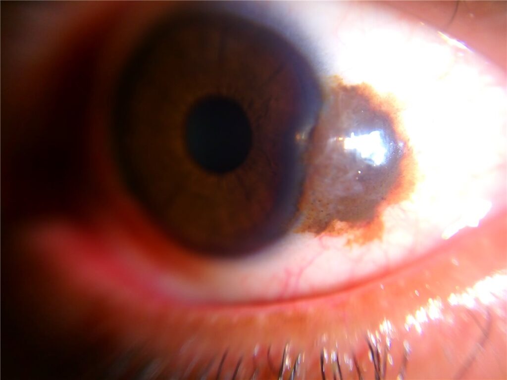
A conjunctival nevus most commonly appears near the limbus, though it can occur anywhere on the bulbar conjunctiva, and less often on the caruncle, plica semilunaris, or, uncommonly, the palpebral conjunctiva.
Pigmentation is variable and this variability is itself a normal feature rather than a warning sign: nevi range from heavily pigmented to amelanotic, and the same lesion can change in apparent pigmentation over time, particularly around puberty or pregnancy, without this representing malignant transformation.
The single most reassuring feature on slit-lamp examination is the presence of clear cystic spaces within the lesion, corresponding to entrapped conjunctival epithelial inclusions. These cysts are a hallmark of benignity and are rarely seen in melanoma, which makes their presence genuinely useful at the bedside rather than just a textbook point.
A benign nevus is typically well circumscribed with distinct borders, mobile over the underlying sclera when the conjunctiva is gently moved with a cotton bud, and does not involve the cornea, though it can abut the limbus closely.
Feeder vessels can be present in benign nevi and are not, on their own, a sign of malignancy, though prominent or newly developed feeder vessels in a lesion that is otherwise changing should prompt closer scrutiny of the whole picture rather than being dismissed automatically.
All-fit Slit-Lamp Adapter
Record and share exactly what you see at the slit lamp. One adapter fits any slit lamp or surgical microscope — and any smartphone.
From Choroida — the team behind this siteDifferential Diagnosis
- Conjunctival melanoma, typically less well circumscribed, often lacking cysts, and more likely to show recent growth, increasing vascularity, or the absence of a clear pre-existing nevus at the same site
- Primary acquired melanosis, a flat, patchy, poorly defined area of pigmentation without the discrete, elevated, cyst-containing appearance of a nevus, and the lesion with the most direct relevance to melanoma risk when atypia is present on biopsy
- Racial or complexion-associated melanosis, typically bilateral, flat, and present since childhood, most pronounced near the limbus, without the discrete raised quality of a nevus
- Conjunctival naevus of Ota or ocular melanocytosis, involving deeper episcleral pigmentation with a slate-grey rather than brown colour, and carrying its own distinct, if still low, malignant potential
- Foreign body or subconjunctival haemorrhage in cases of acute pigmented or discoloured appearance, generally distinguished easily by history and the absence of a discrete mass
Evaluation
Diagnosis is usually made clinically at the slit lamp based on the features described above.
Documentation matters more than any single examination: careful description or, ideally, clinical photography at baseline gives a genuine comparison point for future visits and is often more reliable than a patient’s or clinician’s memory of “how it looked before” at a follow-up appointment months or years later.
Anterior segment OCT can help characterise lesion thickness and internal structure, including the presence of cystic spaces, in cases where the clinical appearance alone leaves some uncertainty.
Biopsy or excision is reserved for lesions with concerning features: documented growth, new or increasing vascularity, loss of cystic spaces in a previously cystic lesion, involvement of the palpebral conjunctiva or caruncle in a way that seems atypical for the patient, or any feature that does not fit the reassuring pattern described above.
Most stable, cyst-containing, well-circumscribed nevi do not need biopsy, and biopsying every pigmented conjunctival lesion on sight would represent significant overtreatment of a generally benign condition.
Management
Observation with periodic clinical examination, typically annually or as clinically indicated, is appropriate for the great majority of conjunctival nevi.
Baseline photography is genuinely useful practice here, since it turns a subjective “does this look different” judgement into an objective comparison at each subsequent visit.
Excision is considered for lesions with atypical or changing features, for significant cosmetic concern, or occasionally for lesions causing mechanical irritation from their size or location.
When excision is performed, a wide margin with adequate tissue processing for histopathology is appropriate, both to confirm the benign diagnosis and to exclude the rare case of an unsuspected melanoma or atypical melanosis found on final pathology.
Prognosis
The overwhelming majority of conjunctival nevi remain benign throughout a patient’s life and never require intervention beyond periodic observation.
Malignant transformation to melanoma is possible but rare, and the risk is not something that should be overstated to patients who otherwise have a textbook benign lesion, since undue anxiety about a well-circumscribed, cystic, stable nevus is rarely warranted.
What does matter is genuine change over time, which is exactly why establishing a documented baseline and following lesions at reasonable intervals, rather than either ignoring pigmented conjunctival lesions entirely or biopsying every one reflexively, is the appropriate long-term approach.


Document what you see
Two smartphone imaging tools built for everyday clinic use — one for the slit lamp, one for the fundus.
From Choroida — the team behind this siteReferences
- Shields CL, Fasiuddin A, Mashayekhi A, Shields JA. Conjunctival nevi: clinical features and natural course in 410 consecutive patients. Archives of Ophthalmology. 2004.
- Shields CL, Shields JA. Tumors of the conjunctiva and cornea. Survey of Ophthalmology. 2004.
- Kenawy N, Lake SL, Coupland SE, Damato BE. Conjunctival melanocytic tumours. Eye. 2013.
- Conjunctival Nevus. EyeWiki, American Academy of Ophthalmology.
- Conjunctival Melanocytic Lesions. StatPearls, NCBI Bookshelf.
Test yourself
A few questions straight from this article.
-
How does conjunctival nevus rank among pigmented conjunctival lesions seen in clinic?
Conjunctival nevus is by far the commonest pigmented conjunctival lesion, usually found incidentally. The great majority need nothing beyond reassurance and periodic monitoring. -
Where does a conjunctival nevus most commonly appear?
Nevi most often sit near the limbus, though they can occur anywhere on the bulbar conjunctiva. The caruncle, plica semilunaris and palpebral conjunctiva are less common sites. -
A conjunctival nevus becomes visibly more pigmented around puberty. How should this be interpreted?
Nevi often become more noticeably pigmented around puberty as hormonal changes affect melanocyte activity, and the same can occur in pregnancy. Patients may perceive this as new growth rather than increased pigmentation of an existing lesion. -
Which histological subtype of conjunctival nevus is most typical in older patients?
Subepithelial nevi lie entirely beneath the epithelium and are more typical of older patients. This reflects nevi migrating deeper into the tissue over time, while junctional nevi predominate in the young. -
Which slit-lamp feature is the single most reassuring sign that a pigmented conjunctival lesion is benign?
Cystic spaces correspond to entrapped conjunctival epithelial inclusions and are a hallmark of benignity, rarely seen in melanoma. Their presence is genuinely useful at the bedside rather than just a textbook point. -
Which examination findings fit a typical benign conjunctival nevus?
A benign nevus has distinct borders and moves over the underlying sclera when the conjunctiva is nudged with a cotton bud. It does not involve the cornea, although it may abut the limbus closely. -
How should feeder vessels in an otherwise typical conjunctival nevus be interpreted?
Feeder vessels appear in benign nevi and are not by themselves a sign of malignancy. Prominent or newly developed vessels in a lesion that is otherwise changing do warrant closer scrutiny of the whole picture. -
Which appearance distinguishes primary acquired melanosis from a conjunctival nevus?
Primary acquired melanosis is flat and poorly demarcated, lacking the discrete, elevated, cyst-containing quality of a nevus. It is the lesion with the most direct relevance to melanoma risk when biopsy shows atypia. -
Which change in a previously typical conjunctival nevus is an indication for biopsy or excision?
Losing cystic spaces in a lesion that previously had them is a concerning feature, alongside documented growth and new or increasing vascularity. Stable, cyst-containing, well-circumscribed nevi do not need biopsy. -
What is the appropriate long-term approach for a stable, well-circumscribed conjunctival nevus?
Observation, typically annually, with baseline photography turns a subjective impression of change into an objective comparison. Malignant transformation is possible but rare, and should not be overstated to a patient with a textbook benign lesion.

Writes and reviews the clinical articles on Educate. Every post is checked against peer-reviewed sources before it goes live.


