
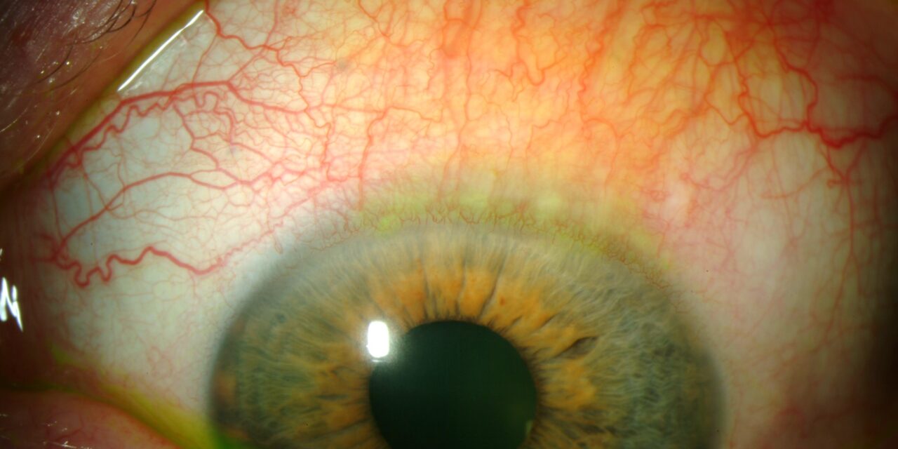
Superior limbic keratoconjunctivitis (SLK) is a chronic inflammatory condition confined to the superior bulbar conjunctiva, superior limbus, and adjacent superior cornea.
It is frequently misdiagnosed because standard treatments for more common causes of red eye simply do not work on it, a clue that, in hindsight, is often the first thing that should raise suspicion for SLK rather than an ordinary conjunctivitis.
The superior location of the disease is itself diagnostic, because most other forms of conjunctivitis and keratitis are not so specifically confined to this one region.
Association with Thyroid Disease
A substantial proportion of patients with SLK have coexisting thyroid dysfunction, most often Graves’ disease.
The mechanical hypothesis for this association centers on excessive laxity of the superior bulbar conjunctiva relative to the globe, which is thought to increase friction between the lid and globe with each blink.
Proptosis and lid retraction from thyroid eye disease may further increase this mechanical stress.
Not every patient with SLK has thyroid disease, but the association is strong enough that thyroid function testing is a reasonable part of the workup in any newly diagnosed case.
Pathogenesis
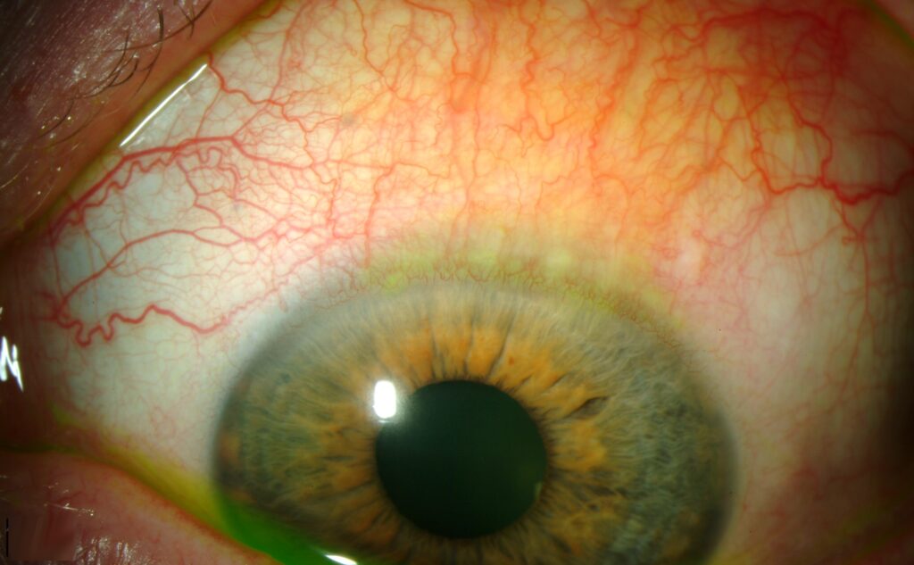
The leading mechanical hypothesis holds that excess, redundant superior bulbar conjunctiva rubs against the upper eyelid with each blink, causing chronic mechanical trauma that in turn produces the characteristic inflammatory changes.
Dry eye and reduced tear film stability appear to be contributing rather than sole causative factors.
Contact lens wear has also been associated with a similar clinical picture, suggesting a shared final mechanical pathway across several different triggers.
This convergence of multiple possible triggers on a single, shared mechanical endpoint is a useful way to think about SLK generally: whatever the initiating factor, the disease behaves and responds to treatment in largely the same way once the characteristic superior conjunctival redundancy and chronic friction pattern are established.
All-fit Slit-Lamp Adapter
Record and share exactly what you see at the slit lamp. One adapter fits any slit lamp or surgical microscope — and any smartphone.
From Choroida — the team behind this siteClinical Presentation
Patients report a foreign-body sensation, burning, and mild photophobia disproportionately localized to the superior aspect of the eye.
Symptoms are frequently bilateral, though sometimes asymmetric, and notoriously chronic and relapsing, sometimes persisting for years despite trials of conventional dry eye and allergy treatments.
The chronic, treatment-resistant nature of the symptoms is itself a diagnostic clue that should prompt a specific, deliberate exam of the superior conjunctiva rather than continuing to treat empirically.
Exam Findings
- Injection and thickening confined to the superior bulbar conjunctiva
- Fine papillary reaction on the superior tarsal conjunctiva
- Superior corneal punctate staining and, in more severe cases, filamentary keratitis
- Redundant, loose superior bulbar conjunctiva, sometimes forming visible folds
- A positive “lid-pull” or superior conjunctival elevation sign, where gently pulling the upper lid up and away from the globe accentuates the injection and folds
Because the affected area sits under the upper lid, SLK is easily missed unless the examiner specifically lifts the lid and directs attention to the superior conjunctiva and limbus.
A routine anterior segment exam that stays focused on the visible palpebral aperture will miss it entirely.
Differential Diagnosis
- Vernal keratoconjunctivitis — giant tarsal papillae and limbal Horner-Trantas dots, typically more diffusely distributed rather than confined superiorly, and with a different age and seasonal pattern
- Giant papillary conjunctivitis — tarsal papillae related to contact lens or foreign body, without the same superior bulbar conjunctival redundancy and thickening
- Thyroid eye disease alone, without true SLK — exposure-related surface changes from proptosis and lid retraction rather than the specific mechanical friction pattern of SLK
- Filamentary keratitis from other causes — dry eye, herpes zoster, or post-surgical, without the characteristic superior limbic distribution
Diagnostic Evaluation
The diagnosis is made clinically, based on the characteristic superior distribution of injection, thickening, and staining, identified by deliberately everting or elevating the upper lid to inspect the superior bulbar conjunctiva and limbus.
Rose bengal or lissamine green staining highlights devitalized superior conjunctival and corneal cells with particular sensitivity in SLK, often more clearly than fluorescein alone, and is a useful adjunct when the diagnosis is uncertain on white-light exam.
Given the association described above, thyroid function testing is a reasonable addition to the initial workup, particularly in a patient without an already established thyroid diagnosis, and identifying previously unrecognized thyroid disease in this context can have significant health implications well beyond the eye finding itself.
Management
Aggressive lubrication is first-line and helps a subset of patients, though many cases are relatively resistant to lubrication alone given the underlying mechanical component.
Silver nitrate application (a dilute solution applied to the affected conjunctiva) is a classic, still frequently used treatment that chemically cauterizes the redundant tissue and can produce significant symptomatic improvement, though it needs to be applied carefully by an experienced clinician given the risk of corneal toxicity if concentration or technique is off.
Bandage contact lenses reduce the mechanical friction between the lid and the redundant conjunctiva and can be effective, for patients who cannot tolerate or do not respond to silver nitrate.
For refractory cases, surgical resection of the redundant superior bulbar conjunctiva (conjunctival recession or excision) directly addresses the excess tissue thought to drive the mechanical irritation, and is reserved for cases that fail these less invasive measures, with topical cyclosporine also reported as a useful adjunct in some chronic, relapsing cases.


Document what you see
Two smartphone imaging tools built for everyday clinic use — one for the slit lamp, one for the fundus.
From Choroida — the team behind this siteReferences
- Theodore FH. Superior limbic keratoconjunctivitis. Eye, Ear, Nose and Throat Monthly.
- Nelson JD. Superior limbic keratoconjunctivitis (SLK). Eye.
- Sahin A, Bozkurt B, Irkec M. Topical cyclosporine A in the treatment of superior limbic keratoconjunctivitis. Cornea.
- American Academy of Ophthalmology. Basic and Clinical Science Course, Section 8: External Disease and Cornea.
Test yourself
A few questions straight from this article.
-
To which region is superior limbic keratoconjunctivitis confined?
SLK is confined to the superior bulbar conjunctiva, superior limbus and adjacent superior cornea, and that strictly superior location is itself diagnostic. -
Which systemic condition most often accompanies superior limbic keratoconjunctivitis?
A substantial proportion of SLK patients have thyroid dysfunction, most often Graves' disease, so thyroid function testing belongs in the workup of any new case. -
What does the leading mechanical hypothesis propose as the cause of SLK?
Excess, lax superior bulbar conjunctiva rubs against the upper lid on blinking, and that chronic mechanical trauma produces the inflammatory changes; dry eye contributes rather than causes. -
Which symptom pattern should raise suspicion for superior limbic keratoconjunctivitis?
Symptoms are chronic, relapsing and localized superiorly, and their failure to respond to conventional dry eye and allergy therapy is often the first real clue to the diagnosis. -
What is the lid-pull sign in superior limbic keratoconjunctivitis?
Gently pulling the upper lid up and away from the globe accentuates the injection and redundant folds; without lifting the lid the disease is missed entirely. -
Which stain best shows the superior conjunctival changes of SLK?
Rose bengal and lissamine green highlight devitalized superior conjunctival and corneal cells with particular sensitivity, often more clearly than fluorescein alone. -
What distinguishes vernal keratoconjunctivitis from superior limbic keratoconjunctivitis?
Vernal disease shows giant tarsal papillae and Horner-Trantas dots distributed more diffusely, with a different age and seasonal pattern, rather than the strictly superior involvement of SLK. -
What is first-line treatment for superior limbic keratoconjunctivitis?
Lubrication is first-line and helps a subset of patients, though many cases resist it because the underlying problem is mechanical rather than purely tear-related. -
How does silver nitrate application help in superior limbic keratoconjunctivitis?
Dilute silver nitrate cauterizes the redundant tissue and can give marked symptomatic relief, but needs careful application by an experienced clinician because of corneal toxicity risk. -
What is reserved for SLK that fails lubrication, silver nitrate and bandage lenses?
Conjunctival recession or excision removes the excess tissue driving the mechanical irritation and is kept for refractory cases; topical cyclosporine is also reported as a useful adjunct.
