
CASE REPORT
A 39-years-old man presented to a clinic with complaints of decreased blurred vision and floaters in his left eye over the previous two weeks.

His best-corrected visual acuity at the initial presentation was 20/ 20 in the right eye and hand movement in the left eye. The anterior segment and intraocular pressure were normal.
The visual field using the Goldmann perimeter and the Humphrey static achromatic automatic perimetry (central 30-2 threshold test) was normally at the right eye and could not be performed on the left eye owing to the markedly decreased visual acuity.
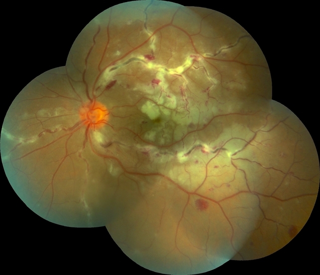
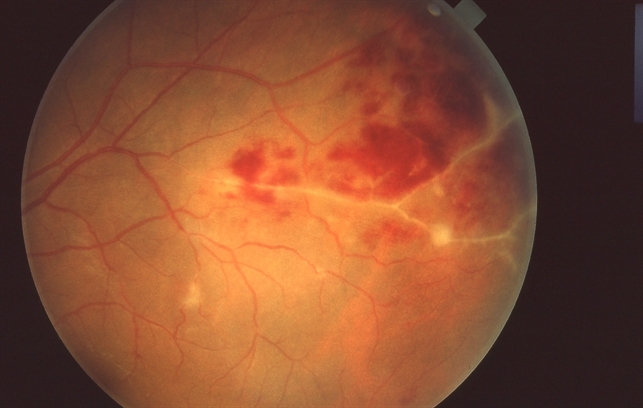
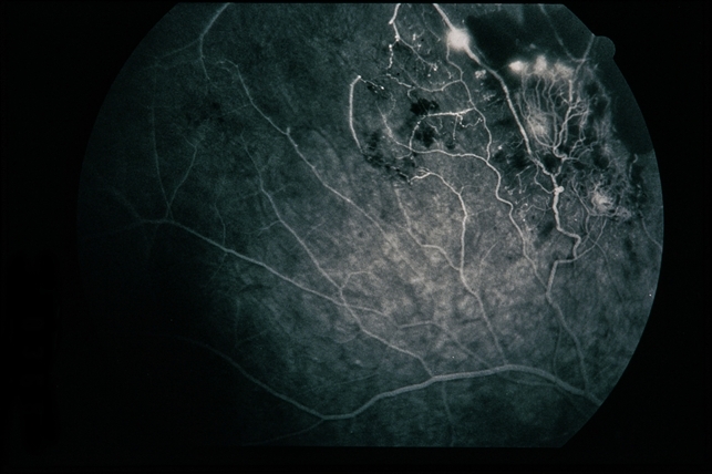
Ultrasonography of the left eye highlighted dense vitreous floaters, which obscured the view of the fundus. Dilated ocular fundus examination of the right eye showed pallor of the optic nerve head; temporally from the macula in the mid-periphery, there were perivascular sheathing, scattered wet perivascular exudates, dotted and flame-shaped intraretinal hemorrhages, focal occlusions of retinal vessels, extensive nonperfused areas of the capillaries from the macula in the mid-retina periphery, which extended towards the posterior pole and retinal periphery, giving rise to preretinal neovascularization prominent into the vitreous in the fashion of a sea fan.
Taking into account all the clinical, angiographic, and ultrasonographic assessments, the diagnoses of bilateral Eales disease in the late (proliferative) stage with neovascularization and retinal and vitreous hemorrhages were established.
DISEASE
Eales’ disease was first described by British ophthalmologist Henry Eales in 1880. Eales’ disease is an idiopathic occlusive vasculitis involving the mid-peripheral retina that is characterized by retinal venous inflammation (periphlebitis), vascular occlusion, and subsequent retinal neovascularization.
A hallmark of Eales’ disease is recurrent vitreous hemorrhage.
Eales’ disease mainly affects young males in their second decade of life. It is more prevalent in India and Middle Eastern countries with cases observed worldwide.
In a study by Biwas et al., a male-to-female ratio of 20:1 was observed with a mean age at presentation of 29.9 years, with a range from 11– 59 years.
MANAGEMENT
General Treatment
The management of Eales’ disease depends upon the stage of the disease. The treatment strategy consists of medical treatment with oral corticosteroids in the active inflammatory stage and laser photocoagulation for retinal ischemia and neovascularization.
Biwas et al. observed that the timely use of oral corticosteroid during active inflammation and the use of laser ablation of areas with capillary non-perfusion had a statistically significant beneficial impact on visual outcomes during a 10-year follow-up period.
Medical Treatment
- Corticosteroids
- Anti-Vascular Endothelial Growth Factor (Anti-VEGF)
- Anti-Tuberculosis Treatment
- Photocoagulation
- Surgical treatment
Would you have interest in taking retina images by smartphone?
Fundus photography is superior to fundus analysis as it enables intraocular pathologies to be photo captured and encrypted information to be shared with colleagues and patients.
Recent technologies allow smartphone-based attachments and integrated lens adaptors to transform the smartphone into a portable fundus camera and Retinal imaging by smartphone.
RETINAL IMAGING BY YOUR SMARTPHONE
REFERENCES
- Eales H. Primary retinal hemorrhage in young men. Ophthalmic Rev. 1882;1: 41.
- Biswas J, Ravi RK, Naryanasamy A, Kulandai LT, Madhavan HN. Eales’ disease – current concepts in diagnosis and management. J Ophthalmic Inflamm Infect. 2013;3(1):11.
- Das T, Pathengay A, Hussain N, Biswas J. Eales’ disease: diagnosis and management. Eye (Lond). 2010;24(3):472-82.
- Biswas J, K R R, Pal B, Gondhale HP, Kharel Sitaula R. Long-Term Outcomes of a Large Cohort of Patients with Eales’ Disease. Ocul Immunol Inflamm. 2018;26(6):870‐876.
- Ishaq M, Karamat S, Niazi MK. HLA typing in patients of Eales disease. J Coll Physicians Surg Pak. 2005 May; 15(5):288-90.
- Rajesh M, Sulochana KN, Coral K, et al. Determination of carbonyl group content in plasma proteins as a useful marker to assess impairment in antioxidant defense in patients with Eales’ disease. Indian J Ophthalmol. 2004;52(2):139-144.
