
Argon laser peripheral iridoplasty uses low-energy laser burns applied to the peripheral iris to contract and physically pull the iris root away from the trabecular meshwork, addressing angle closure that persists despite a patent peripheral iridotomy, discussed in its own dedicated article on this site.
It works through an entirely different mechanism than iridotomy: rather than relieving pupillary block by creating an alternate aqueous pathway, iridoplasty mechanically reshapes and flattens the peripheral iris configuration itself, making it the specific tool for angle closure driven by anatomy rather than by pupillary block.
Understanding this mechanistic difference clarifies why iridoplasty is reserved for a fairly specific subset of angle-closure presentations, mainly plateau iris syndrome, rather than being used as a general angle-closure treatment interchangeable with iridotomy.
The procedure is used considerably less often than iridotomy in everyday practice, reflecting how much less common plateau iris configuration is compared to straightforward pupillary block as a cause of angle closure overall.
Mechanism
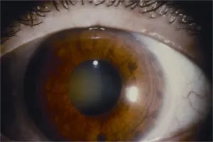
Low-energy, relatively large-spot laser burns applied to the peripheral iris cause localized tissue contraction, physically pulling and flattening the treated area of iris away from the adjacent trabecular meshwork and angle structures.
This mechanical effect is fundamentally different from iridotomy’s function, which relieves a pressure differential between the posterior and anterior chambers rather than physically reshaping the iris configuration itself.
Because the effect works by directly altering iris configuration and angle anatomy, iridoplasty is specifically suited to angle closure where the anatomic configuration of the peripheral iris, rather than pupillary block, is the primary mechanism causing angle crowding.
Plateau Iris Syndrome
In plateau iris configuration, the peripheral iris sits abnormally forward due to an anteriorly positioned ciliary body, crowding the angle even when pupillary block has already been fully relieved by a patent iridotomy.
Plateau iris syndrome describes this same anatomic configuration when it produces symptomatic or measurable angle closure, and it is the classic and most well-established indication for iridoplasty, since the mechanism, anatomic crowding by the iris root itself, matches exactly what iridoplasty is designed to correct.
Recognizing that an iridotomy alone will not resolve angle closure driven by plateau iris configuration is essential, since continuing to rely on iridotomy alone in this specific scenario leaves the underlying anatomic problem unaddressed.
Ultrasound biomicroscopy, discussed in relation to anterior segment imaging elsewhere on this site, is particularly useful for confirming a plateau iris configuration directly, since it can visualize the anteriorly positioned ciliary processes that standard anterior segment OCT cannot see behind the iris.
All-fit Slit-Lamp Adapter
Record and share exactly what you see at the slit lamp. One adapter fits any slit lamp or surgical microscope — and any smartphone.
From Choroida — the team behind this siteOther Indications
- Acute angle-closure attack unresponsive to initial medical therapy, where iridoplasty can be used as an emergency measure to mechanically open the angle and break the acute attack while more definitive treatment is arranged
- Angle closure from a lens-related mechanism where the peripheral iris configuration continues contributing to angle crowding despite iridotomy
- Adjunctive use before or during laser iridotomy in an eye with a very shallow, difficult-to-access anterior chamber, where iridoplasty can temporarily deepen the chamber and facilitate safer iridotomy placement
Technique
The procedure is performed at the slit lamp using a gonioscopy-style contact lens, with low-energy, relatively large, long-duration laser spots applied circumferentially around the peripheral iris.
Energy settings are deliberately kept low, and spot size relatively large, distinguishing the technique from the higher-energy, smaller-spot settings used for iridotomy, since the goal here is gentle tissue contraction rather than creating a full-thickness perforation.
The procedure typically requires treatment around most or all of the iris circumference to achieve a meaningful, sustained widening effect on the angle.
Limitations and Complications
The effect of iridoplasty can diminish over time in some patients, sometimes requiring repeat treatment as the angle configuration gradually reverts toward its original, more crowded anatomy.
Transient intraocular pressure elevation, mild anterior chamber inflammation, and localized iris pigment changes at the treatment site are recognized, generally self-limited effects of the procedure.
Iridoplasty does not replace iridotomy when pupillary block is present, and it does not address the underlying anatomic predisposition permanently in the way that lens extraction can in an eye where the lens itself is a significant contributor to angle crowding.
For this reason, iridoplasty is often best understood as one component within a broader, individualized angle-closure management plan rather than a single definitive solution on its own.


Document what you see
Two smartphone imaging tools built for everyday clinic use — one for the slit lamp, one for the fundus.
From Choroida — the team behind this siteReferences
- Ritch R, Tham CC, Lam DS. Argon laser peripheral iridoplasty (ALPI): an update. Survey of Ophthalmology.
- American Academy of Ophthalmology. Basic and Clinical Science Course, Section 10: Glaucoma.
- Ritch R. Plateau iris is caused by abnormally positioned ciliary processes. Journal of Glaucoma.
Test yourself
A few questions straight from this article.
-
How do argon laser burns widen the angle in peripheral iridoplasty?
Low-energy burns cause localised contraction of the peripheral iris, physically pulling and flattening it away from the trabecular meshwork and adjacent angle structures. -
How does iridoplasty differ mechanistically from laser peripheral iridotomy?
Iridotomy relieves pupillary block by creating an alternate aqueous pathway. Iridoplasty instead mechanically reshapes and flattens the peripheral iris itself. -
Which condition is the classic and best-established indication for argon laser iridoplasty?
Plateau iris syndrome is the classic indication because its mechanism, anatomic crowding by the iris root itself, matches exactly what iridoplasty is designed to correct. -
What anatomic abnormality underlies plateau iris configuration?
In plateau iris configuration the peripheral iris sits abnormally forward because of an anteriorly positioned ciliary body, crowding the angle even after pupillary block has been relieved. -
Which imaging modality best confirms plateau iris configuration directly?
Ultrasound biomicroscopy can visualise the anteriorly positioned ciliary processes behind the iris, which standard anterior segment optical coherence tomography cannot see. -
In which acute situation can iridoplasty be used as an emergency measure?
When an acute attack does not respond to initial medical therapy, iridoplasty can mechanically open the angle and break the attack while more definitive treatment is arranged. -
How can iridoplasty assist when the anterior chamber is very shallow and hard to access?
In an eye with a very shallow, difficult-to-access anterior chamber, iridoplasty can temporarily deepen the chamber and make iridotomy placement safer. -
Which laser settings characterise argon laser peripheral iridoplasty?
Energy is kept deliberately low and spot size relatively large, unlike the higher-energy, smaller spots used for iridotomy, because the aim is gentle contraction rather than perforation. -
Which side effects are recognised after argon laser peripheral iridoplasty?
Transient intraocular pressure elevation, mild anterior chamber inflammation and localised iris pigment changes at the treatment site are recognised and generally self-limited. -
What limitation should be discussed before performing argon laser peripheral iridoplasty?
The effect can fade in some patients as the angle configuration gradually reverts toward its original crowded anatomy, sometimes requiring repeat treatment.
