
Progressive outer retinal necrosis (PORN) is a rapidly progressive necrotizing retinitis caused mainly by varicella zoster virus in patients with profound immunosuppression.
Unlike acute retinal necrosis, it produces little or no vitritis and no retinal vasculitis, so the eye can look deceptively quiet while vision is being lost.
Without prompt treatment the necrosis spreads over days and often leads to retinal detachment.
Recognition rests on the appearance of the fundus and on the immune status of the patient.
What Is Progressive Outer Retinal Necrosis?
PORN was first described in patients with AIDS, and it typically occurs when the CD4 count is very low, often below 50 cells/µL.
It can also occur in other severely immunocompromised patients, such as recipients of organ or stem cell transplants and patients on heavy immunosuppression.
Varicella zoster virus is the usual cause, and herpes simplex virus is reported less often.
Many patients have a history of herpes zoster, sometimes with cutaneous or neurologic involvement, before or around the time of retinitis.
The disease differs from acute retinal necrosis (ARN), which occurs in immunocompetent or mildly immunosuppressed patients and features prominent vitritis and vasculitis (see acute retinal necrosis).
Clinical Features
Patients complain of blurred vision, floaters, or visual field loss, and the symptoms are often painless.
The anterior chamber is usually quiet, and vitreous cells are minimal.
Fundus findings include:
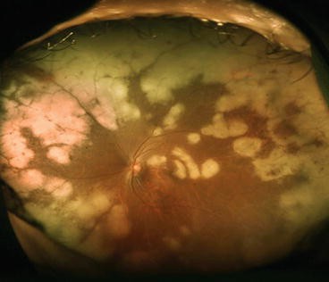
- Multifocal patches of deep retinal whitening that begin in the outer retina
- Early involvement of the posterior pole and macula, as well as the periphery
- Lesions that coalesce rapidly into a large area of full-thickness necrosis
- A characteristic cracked-mud appearance in the affected retina
- Relative sparing of retinal vessels, with little or no perivascular sheathing
Necrotic areas soon become atrophic, and retinal breaks develop within weeks.
Retinal detachment is common and often bilateral, and it is frequently complicated by proliferative vitreoretinopathy.
The disease can progress in a few days and can involve the second eye rapidly.
Fundus Explorer Pro
Photograph the retinal findings described here with the phone already in your pocket — 22 D optics and built-in illumination in one handheld unit.
From Choroida — the team behind this siteWhy the Eye Looks Quiet
The lack of inflammation reflects the patient’s immune status.
Without functioning CD4 T cells, the eye cannot mount the vitreous and vascular response that characterizes ARN, so the virus spreads through the outer retina largely unopposed.
The outer retina is involved first because varicella zoster virus enters through the retinal pigment epithelium and photoreceptor layer and spreads laterally.
This explains the deep, cloud-like whitening and the absence of vascular sheathing.
The same absence of inflammation can delay diagnosis, since a quiet anterior chamber and clear vitreous may falsely reassure the examiner.
Patients often present with a large area of necrosis that has developed over a few days.
Differential Diagnosis
Distinguishing PORN from other necrotizing retinitis matters because treatment and prognosis differ.
- CMV retinitis progresses more slowly and produces a granular border with hemorrhage along the vessels (see CMV retinitis)
- Acute retinal necrosis shows marked vitritis, peripheral necrosis, and occlusive arteritis
- Ocular toxoplasmosis causes a focal retinochoroiditis with overlying vitritis, often next to an old scar
- Syphilitic retinitis may resemble PORN and should be tested for in every patient
- Intraocular lymphoma may cause retinal infiltrates in immunosuppressed patients
Many patients have coinfections, so testing for other opportunistic infections is appropriate.
Diagnosis
The diagnosis is clinical, and it is confirmed by PCR of aqueous or vitreous fluid for varicella zoster virus, herpes simplex virus, and cytomegalovirus.
Aqueous sampling is quick and adequate in most cases, and vitreous sampling is used when the aqueous result is negative but suspicion remains high.
Evaluation should include CD4 count, HIV viral load in HIV-positive patients, and syphilis serology.
Fundus photography and widefield imaging document the extent of disease and help track progression.
OCT of the macula shows outer retinal hyperreflectivity and loss of normal layers in affected areas.
Treatment
Treatment should start as soon as the diagnosis is suspected, without waiting for laboratory confirmation.
Antiviral Therapy
The most widely used approach combines intravenous antiviral drugs (ganciclovir, foscarnet, or both) with repeated intravitreal injections of ganciclovir or foscarnet.
The combination is used because monotherapy has been associated with poor outcomes in published series.
Acyclovir alone is not considered adequate for PORN.
Treatment usually continues for several weeks, and it is followed by long-term maintenance therapy until immune function recovers.
Immune Reconstitution
In HIV-infected patients, antiretroviral therapy should be started or optimized in coordination with the infectious disease team.
Immune recovery can improve the long-term outlook, but it may also cause inflammation, so patients need careful follow-up.
Detachment and Surgery
Prophylactic laser demarcation of the border of viable retina has been described, although evidence is limited.
Retinal detachment is managed with vitrectomy and long-acting tamponade, usually silicone oil, since PVR is frequent and gas tamponade often fails.
Practical Points at the Bedside
- Examine both eyes carefully with a wide view, since early lesions may be small and peripheral
- Ask about recent zoster, varicella exposure, headache, and neurologic symptoms
- Send aqueous for PCR at the first visit, and do not delay antiviral therapy for the result
- Arrange baseline photographs or widefield imaging to measure progression between visits
- Check renal function before intravenous ganciclovir or foscarnet, and watch for marrow toxicity and electrolyte disturbance
- Involve the infectious disease team early and review the patient within one to two days
Intravitreal injections are usually repeated twice weekly at first, then tapered as the lesions stabilize.
Systemic foscarnet needs hydration and monitoring of calcium, magnesium, and creatinine.
Patients should be told that the second eye is at risk and should report any new visual symptoms immediately.
Prognosis
The visual outcome remains poor in many patients, and no light perception in the affected eye is common in older series.
Outcomes are better with early recognition, combination antiviral therapy, and immune recovery.
Ophthalmologists who care for immunocompromised patients should be aware that a quiet eye with rapidly spreading retinal whitening is PORN until proven otherwise.


Document what you see
Two smartphone imaging tools built for everyday clinic use — one for the slit lamp, one for the fundus.
From Choroida — the team behind this siteReferences
- Forster DJ, Dugel PU, Frangieh GT, Liggett PE, Rao NA. Rapidly progressive outer retinal necrosis in the acquired immunodeficiency syndrome. Am J Ophthalmol. 1990;110:341-348.
- Margolis TP, Lowder CY, Holland GN, et al. Varicella-zoster virus retinitis in patients with the acquired immunodeficiency syndrome. Am J Ophthalmol. 1991;112:119-131.
- Engstrom RE Jr, Holland GN, Margolis TP, et al. The progressive outer retinal necrosis syndrome: a variant of necrotizing herpetic retinopathy in patients with AIDS. Ophthalmology. 1994;101:1488-1502.
- Holland GN. Standard diagnostic criteria for the acute retinal necrosis syndrome. Executive Committee of the American Uveitis Society. Am J Ophthalmol. 1994;117:663-667.
- Schoenberger SD, Kim SJ, Thorne JE, et al. Diagnosis and treatment of acute retinal necrosis: a report by the American Academy of Ophthalmology. Ophthalmology. 2017;124:382-392.
