
Orbital lymphangioma is a benign but often clinically challenging vascular malformation composed of abnormal lymphatic (and frequently intermixed venous) channels, distinguished from infantile capillary hemangioma by two features with major practical consequences: it typically does not proliferate and involute the way a true infantile hemangioma does, and it has a genuine tendency toward sudden, dramatic enlargement from spontaneous intralesional hemorrhage — an event that can transform a previously stable, well-tolerated lesion into an acute orbital emergency within hours.
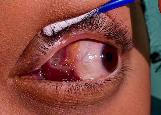
Nature of the Lesion
Orbital lymphangiomas are congenital, low-flow vascular malformations rather than true neoplasms, generally classified as part of the broader spectrum of vascular anomalies that includes both purely lymphatic and combined venolymphatic malformations.
Unlike infantile capillary hemangioma, which reliably proliferates in the first year of life and then involutes over subsequent years, lymphangioma tends to grow proportionately with the child (or remain stable) rather than following that same predictable proliferation-involution trajectory, making its natural history considerably less predictable and less reassuring to families than that of a typical capillary hemangioma.
Clinical Presentation
Presentation is highly variable: some lymphangiomas are relatively quiescent for years before an acute event brings them to attention, while others produce chronic, slowly progressive proptosis and eyelid or conjunctival swelling from the outset.
Growth often becomes apparent or accelerates during upper respiratory infections (from associated lymphoid tissue enlargement) or, more dramatically, from spontaneous intralesional hemorrhage into one of the malformation’s abnormal channels, producing the sudden, marked swelling sometimes described as a “chocolate cyst” once the hemorrhage has organized within the lesion.
All-fit Slit-Lamp Adapter
Record and share exactly what you see at the slit lamp. One adapter fits any slit lamp or surgical microscope — and any smartphone.
From Choroida — the team behind this siteThe Hemorrhage Problem
Spontaneous hemorrhage into a lymphangioma can occur with little or no warning, causing acute, sometimes severe proptosis, pain, and, in significant cases, compressive optic neuropathy from the sudden increase in orbital volume.
This acute hemorrhagic presentation is the single most clinically urgent aspect of managing this condition, and families of children with a known orbital lymphangioma should be counseled about the possibility in advance, because recognizing it promptly (rather than assuming a slower-growing process) directly affects how quickly appropriate emergency care is sought.
Recurrent hemorrhage into the same lesion is common, and each episode carries its own risk to vision depending on severity and how promptly it is recognized and treated, which is why ongoing vigilance is needed even after a first episode has resolved without lasting harm.
Exam and Imaging Findings
- Superficial conjunctival or eyelid components may appear as small, clear or blood-tinged cystic vesicles, sometimes described as having a “bag of worms” quality on palpation when the lesion is more extensive
- Proptosis and globe displacement with deeper orbital involvement, variable in degree and sometimes fluctuating over time with the lesion’s hemorrhagic episodes
- MRI is the imaging study of choice, showing a poorly circumscribed, multiloculated lesion with characteristic fluid-fluid levels when hemorrhage has occurred into some of the cystic spaces — a finding that is genuinely useful for confirming the diagnosis and distinguishing it from other orbital masses
- Signs of optic nerve compression (reduced vision, a relative afferent pupillary defect, discussed in more detail in this site’s dedicated article on that sign) in cases with significant acute hemorrhage or chronic mass effect
Differential Diagnosis
- Infantile capillary hemangioma — proliferates and reliably involutes, without the same tendency toward sudden hemorrhagic enlargement, and with a different, more homogeneous imaging appearance, discussed in its own dedicated article on this site
- Orbital dermoid cyst — a stable, non-hemorrhagic, well-circumscribed lesion with an entirely different natural history and imaging appearance, discussed in its own dedicated article on this site
- Rhabdomyosarcoma — a more rapidly and progressively enlarging malignant mass, an important consideration to exclude, particularly when the presentation is atypical for the more classic, fluctuating course of lymphangioma
- Orbital varix — also shows positional or Valsalva-related size change, but through a different, purely venous mechanism, discussed in its own dedicated article on this site, and generally without the same fluid-fluid level pattern on imaging
Management
Small, stable, asymptomatic lymphangiomas can be observed, with clear counseling to the family about the possibility of future acute hemorrhagic enlargement and what to watch for.
Acute hemorrhage causing significant proptosis or threatening vision may require urgent intervention: needle aspiration or drainage of the hemorrhagic cyst, or, in severe cases with optic nerve compression, more urgent surgical decompression, generally reserved for vision-threatening presentations rather than used routinely for every hemorrhagic episode.
For lesions requiring more definitive treatment given recurrent hemorrhage, significant cosmetic concern, or progressive growth, options include sclerotherapy (injecting an agent into the abnormal channels to induce scarring and shrinkage) and surgical debulking, though complete surgical excision is often difficult to achieve given how extensively the malformation typically infiltrates and intermixes with normal orbital tissue.
This infiltrative quality is a genuine limitation of surgery as a curative option and is why staged, more conservative management (observation punctuated by intervention only for symptomatic episodes) is frequently the practical, real-world approach rather than attempting a single definitive cure.
Families benefit from understanding this reality from the outset, since expecting a single surgical procedure to fully resolve the condition often does not match the actual, longer-term course most patients experience.


Document what you see
Two smartphone imaging tools built for everyday clinic use — one for the slit lamp, one for the fundus.
From Choroida — the team behind this siteReferences
- Wright JE, Sullivan TJ, Garner A, et al. Orbital venous anomalies. Ophthalmology.
- Harris GJ. Orbital vascular malformations: a consensus statement on terminology and its clinical implications. American Journal of Ophthalmology.
- American Academy of Ophthalmology. Basic and Clinical Science Course, Section 7: Orbit, Eyelids, and Lacrimal System.
