
CASE REPORT
Case report of Choroidal Osteoma 70-year-old male was referred to a clinic with fundoscopic abnormality in the left eye. His visual acuity was 25/20 in both eyes on examination. Slit-lamp examination and intraocular pressures were normal in both eyes.

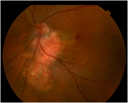
Fundus examination revealed a yellowish–white lesion of 0.9-mm disc diameter in the left eye. OCT scan showed mild sub-RPE elevation corresponding to the yellowish lesion. There was a subtle detachment of the RPE.
The intralesional structure beneath the RPE was unclear. Bruch’s membrane and the sclera-choroidal junction at the lesion were detectable. The choriocapillaris and the inner part of the choroidal vessels were not visible.
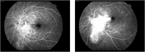
FA revealed staining of the plaque in the late phase of the angiogram, and ICGA revealed hypofluorescence in the early and late phase. B-scan USG in the left eye suspected a thin hypoechoic shadow, but it was not definite for diagnosis.
A thin-slice (1 mm) CT scan of the orbit was performed and showed the lesion had the density of bony tissue, confirming that the diagnosis was choroidal osteoma.
DISEASE of Choroidal Osteoma
It is a benign ossifying tumor characterized by mature bone replacing choroid. The condition is reported rarely, with only 61 patients seen at a major tertiary center in 26 years.
However,
asymptomatic patients with choroidal osteoma are typically not referred to a tertiary center, but rather are followed in community-based practice.
one study reports 11 patients seen in a community-based practice in a 14-year span. This disease is unique as it affects otherwise healthy eyes.
MANAGEMENT of Choroidal Osteoma
Asymptomatic or stable choroidal osteoma can be observed.
For CNVM, surgical removal and PDT have been used in the past, as well as laser treatment. More recently, anti-VEGF treatments such as intravitreal ranibizumab and bevacizumab have been employed with success.
It has been shown that the margins of the choroidal osteoma that are decalcified have no tumor growth and display the stabilization of the tumor scar.
Therefore,
it has been proposed for the calcified extrafoveal osteoma, to use PDT at the edges to decalcify the tumor and prevent its growth and foveal involvement.
would you have the interest to take retina images by smartphone?
Fundus photography is superior to fundus analysis as it enables intraocular pathologies to be photo captured and encrypted information to be shared with colleagues and patients.
Recent technologies allow smartphone-based attachments and integrated lens adaptors to transform the smartphone into a portable fundus camera.

REFERENCES
- Shields CL, Sun H, Demirci H, Shields JA. Factors predictive of tumor growth, tumor decalcification, choroidal neovascularization, and visual outcome in 74 eyes with choroidal osteoma. Arch Ophthalmol 2005;123:1658-1666.
- Browning DJ. Choroidal osteoma: Observations from a community setting. Ophthalmology 2003;110:1327-1334.
- Shields CL, Shields JA, Augsburger JJ. Choroidal osteoma. Surv Ophthalmol 1988;33:17-27.
- Gass JD, Guerry RK, Jack RL, Harris G. Choroidal osteoma. Arch Ophthalmol 1978;96:428-435.
- Williams AT, Font RL, Van Dyk HJ, Riekhof FT. Osseous choristoma of the choroid simulating a choroidal melanoma. association with a positive 32P test. Arch Ophthalmol 1978;96:1874-1877.
Read more: Choroidal Folds, Birdshot Retinochoroidopathy, Tadpole Pupil, Cerulean Cataract, Coats disease.
