
Trichinosis, caused by the nematode Trichinella spiralis acquired from eating undercooked meat containing encysted larvae, produces periorbital and eyelid edema that is often among the earliest and most recognizable clinical signs of systemic infection, appearing before the muscle pain and fever that more classically define the illness in most people’s minds.
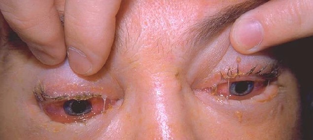
Bilateral periorbital swelling in a patient with a compatible dietary history and evolving systemic symptoms should prompt specific consideration of trichinosis, a diagnosis that is easy to overlook in regions where the infection has become uncommon.
Pathogenesis
After ingestion, larvae are released in the stomach and mature into adults in the small intestine.
The resulting new generation of larvae migrates through the bloodstream to strike striated muscle throughout the body, including the extraocular muscles.
The periorbital tissue swelling seen clinically reflects both direct larval migration into this tissue and, importantly, a hypersensitivity reaction to the parasite that develops as the immune system responds to migrating larvae.
This immune-mediated component is why the periorbital edema is often disproportionate to the number of parasites actually present locally.
The extraocular muscles are a particularly favored site of larval encystment relative to many other striated muscles in the body, likely related to their high metabolic activity and rich blood supply, which is part of why ocular and periocular findings are so often among the presenting features of the disease rather than an incidental, late-appearing complication.
Once encysted within muscle tissue, larvae can remain viable for a prolonged period, eventually calcifying, which is why a history of a prior trichinosis infection can sometimes be inferred incidentally from calcified deposits seen on unrelated imaging performed years later.
Clinical Presentation
- Bilateral periorbital and eyelid edema, often one of the earliest visible signs of systemic infection
- Subconjunctival and retinal hemorrhages, related to the vasculitic component of the immune response to migrating larvae
- Chemosis
- Extraocular muscle pain and, occasionally, restricted eye movement, from larval invasion of the extraocular muscles themselves
- Systemic symptoms developing over the following days — fever, myalgia, and gastrointestinal symptoms from the initial intestinal phase of infection
The combination of periorbital edema with muscle pain, including extraocular muscle involvement causing pain with eye movement, is a distinctive enough pattern that it should prompt specific questioning about recent consumption of undercooked pork, wild game (bear, wild boar), or other meat sources known to harbor the parasite.
Because gastrointestinal symptoms from the initial intestinal phase (nausea, diarrhea, abdominal pain) typically precede the periorbital and muscular findings by several days, a history of a recent, self-limited gastrointestinal illness in someone who now presents with facial swelling and myalgia can be a useful additional clue tying the presentation together.
All-fit Slit-Lamp Adapter
Record and share exactly what you see at the slit lamp. One adapter fits any slit lamp or surgical microscope — and any smartphone.
From Choroida — the team behind this siteDifferential Diagnosis
- Allergic periorbital edema — itchy, related to a clear allergen exposure, without the systemic muscle involvement of trichinosis
- Orbital cellulitis — usually unilateral, with fever, marked pain, and often proptosis or restricted motility from a different mechanism
- Chagas disease (Romana’s sign) — unilateral rather than bilateral periorbital edema, and associated with a specific inoculation site rather than hematogenous larval spread
- Dermatomyositis — periorbital heliotrope discoloration with muscle involvement, but a distinct autoimmune rather than infectious mechanism, and a different systemic symptom pattern and time course
Diagnosis
Eosinophilia is a characteristic and often striking laboratory finding, developing as the larvae migrate through tissue.
Serologic testing (ELISA for Trichinella-specific antibodies) confirms the diagnosis, generally becoming positive a few weeks after infection as the antibody response develops.
Muscle biopsy, once a more central diagnostic tool, is now reserved for cases where serology is inconclusive, given the availability of reliable serologic testing in most clinical settings.
A careful dietary history — asking specifically about undercooked pork or wild game meat consumed in the preceding weeks — is often what points toward the diagnosis in the first place, especially in a patient presenting with the nonspecific combination of periorbital swelling, fever, and muscle pain.
Because serology can remain negative in the earliest days of infection before an antibody response has developed, a single negative test in a patient with a strongly compatible history and exposure should not immediately exclude the diagnosis, and repeat testing or empiric treatment pending confirmation is sometimes reasonable in that setting.
Management
Antiparasitic therapy (albendazole or mebendazole) is most effective when started early in the intestinal phase of infection, before extensive larval migration into muscle tissue has occurred, though treatment is still generally given once the diagnosis is confirmed regardless of the stage, alongside supportive care.
Corticosteroids are added for more severe cases, with significant myositis or evidence of a more generalized hypersensitivity response, to control the inflammatory component of the disease rather than the parasite burden itself.
The periorbital and ocular findings typically resolve as the systemic infection is treated and the acute inflammatory phase settles, generally without leaving lasting ocular sequelae in appropriately treated cases, though severe cases with more extensive muscle involvement can have a longer recovery course overall.
Public health reporting is generally warranted once trichinosis is confirmed, since a single case often signals a shared, potentially contaminated meat source that could affect other people who consumed the same product, making case identification relevant well beyond the individual patient being treated.


Document what you see
Two smartphone imaging tools built for everyday clinic use — one for the slit lamp, one for the fundus.
From Choroida — the team behind this siteReferences
- Gottstein B, Pozio E, Nöckler K. Epidemiology, diagnosis, treatment, and control of trichinellosis. Clinical Microbiology Reviews.
- Dupouy-Camet J, Kociecka W, Bruschi F, et al. Opinion on the diagnosis and treatment of human trichinellosis. Expert Opinion on Pharmacotherapy.
- American Academy of Ophthalmology. Basic and Clinical Science Course, Section 8: External Disease and Cornea.
Test yourself
A few questions straight from this article.
-
Trichinosis is acquired by eating undercooked meat containing what?
The nematode Trichinella spiralis is acquired from undercooked meat carrying encysted larvae, classically pork or wild game such as bear and wild boar. -
Which ocular finding is typically among the earliest visible signs of trichinosis?
Bilateral periorbital and lid oedema is often the earliest and most recognisable sign, appearing before the fever and myalgia that more classically define the illness. -
Why is periorbital oedema in trichinosis often disproportionate to the number of parasites present locally?
The swelling reflects both direct larval migration into periorbital tissue and an immune hypersensitivity reaction, and it is that immune component that makes it disproportionate. -
Why are the extraocular muscles a particularly favoured site of larval encystment in trichinosis?
Their metabolic activity and blood supply make the extraocular muscles a preferred site, which is why ocular and periocular findings so often present early rather than late. -
Subconjunctival and retinal haemorrhages in trichinosis are related to which mechanism?
These haemorrhages reflect the vasculitic element of the host immune response to larvae migrating through tissue. -
What separates Romana's sign of Chagas disease from the periorbital oedema of trichinosis?
Romana's sign is unilateral oedema at an inoculation site, whereas trichinosis produces bilateral swelling from haematogenous larval spread. -
Which laboratory finding is characteristic and often striking in trichinosis?
Eosinophilia is characteristic and often striking, developing as larvae migrate through tissue. -
Trichinella serology is negative in the first days of illness in a patient with strongly compatible exposure. What does this indicate?
ELISA generally turns positive only a few weeks after infection, so an early negative result in a compatible case warrants repeat testing or empiric treatment pending confirmation. -
When is antiparasitic therapy for trichinosis most effective?
Albendazole or mebendazole works best before larvae have migrated extensively into muscle, though treatment is still given once the diagnosis is confirmed at any stage. -
What is the role of corticosteroids in severe trichinosis?
Corticosteroids are added in severe cases with significant myositis or a generalised hypersensitivity response; they address inflammation, not parasite burden.
