
CASE REPORT
A 12-year-old Caucasian male was referred to the Congenital Cataract Section complaining of progressive loss of vision. External observation showed bilateral leukocoria and right exotropia. The best corrected visual acuity was counting fingers at 0.5 m in each eye.

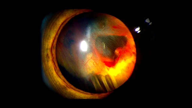
Slit-lamp examination revealed a total cataract in the right eye and a nuclear and posterior subcapsular cataract in the left eye. Goldmann tonometry was 11 mmHg and 10 mmHg, respectively, in the right and left eyes. Right eye fundus examination was not feasible due to total cataracts.
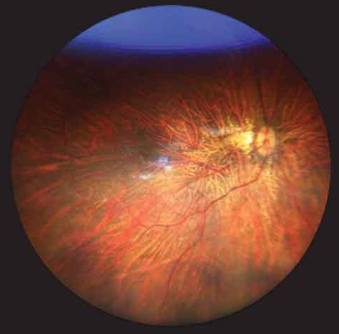
Fundus examination of the left eye showed a myopic fundus with a vitreous detachment and myopic macular scarring. Thin lenses and a probable posterior staphyloma in both eyes were identified by ultrasonography. Vitreous opacities resembling the presence of posterior fetal vasculature were detected in the right eye.
The axial length was 27.8 mm and 27.5 mm, respectively, in the right and left eyes. The patient had no systemic changes or delays in neuro-psychomotor development.
His brother was blind secondary to bilateral retinal detachment and aphakia. Consanguinity was detected in their parents, who were first cousins. A prior familial genetic study had been performed at another institution before the referral. The diagnosis was made as Knobloch syndrome.
Knobloch Syndrome DISEASE entity
Knobloch syndrome is a rare recessively inherited disorder characterized by high myopia, vitreoretinal degeneration, and occipital skull defects, with a high degree of phenotypic variability. First described by Knobloch and Layer in 1971,
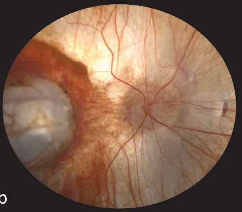
Mutations in the COL18A1 gene are causative for Knobloch syndrome, leading to the production of abnormal type XVIII collagen proteins.
Knobloch syndrome is a rare disorder and since its original report, at least 90 cases from 48 families have been described. Knobloch syndrome has not been reported to be correlated to any specific ethnic group.
Cases have been reported in many ethnic groups including Brazilian, North American, Algerian, Hungarian, El Salvadoran, Saudi Arabian, and Chinese.
Fundus Explorer Pro
Photograph the retinal findings described here with the phone already in your pocket — 22 D optics and built-in illumination in one handheld unit.
From Choroida — the team behind this siteDifferential diagnosis
Differential diagnoses may include other forms of congenital vitreoretinopathies, although each has unique defining features. In Stickler syndrome, there is often radial perivascular retinal degeneration, a lack of macular atrophic lesions, good best-corrected visual acuity, and midface hypoplasia.
Autosomal dominant vitreoretinochoroidopathy shows peripheral and delineated severe retinal degeneration and is often associated with anterior segment dysgenesis.
MANAGEMENT of Knobloch Syndrome
General treatment:
Treatment for Knobloch syndrome is supportive and aimed at treating the symptoms in each individual including surgical repair of both ocular and occipital skull defects.
Ophthalmologic treatment:
Lens extraction may be indicated for ectopia lentis or lenticular opacification, but there is a substantially increased risk of capsular rupture (likely due to modified collagen affecting the integrity of the lens capsule and zonules). Due to the high risk of retinal detachment, prophylactic scleral buckle placement, and/or laser or cryo-retinopexy are sometimes offered.
Genetic planning:
Knobloch syndrome is an autosomal recessive condition that requires a mutation in both copies of the COL18A1 gene. Parents of an affected individual carrying a single mutated copy of this gene do not present with the clinical syndrome but confer a risk of 25% for Knobloch syndrome to each offspring.
Prognosis
Ocular abnormalities in Knobloch syndrome are severe, progressive, and irreversible, usually leading to bilateral blindness. The incidence of retinal detachment is nearly universal even with surgical intervention and prophylactic cryotherapy. Retinal detachments tend to occur at the end of the first decade of life or later.


Document what you see
Two smartphone imaging tools built for everyday clinic use — one for the slit lamp, one for the fundus.
From Choroida — the team behind this siteREFERENCES
- Balikova I, Sanak NS, Fanny D, et al. Three cases of molecularly confirmed Knobloch syndrome. Ophthalmic Genetics 2020;41(1):83-87 doi: 10.1080/13816810.2020.1737948
- Hull S, Arno G, Ku CA, et al. Molecular and Clinical Findings in Patients With Knobloch Syndrome. JAMA Ophthalmology 2016;134(7):753-62 doi: 10.1001/jamaophthalmol.2016.1073
- Zhang LS, Li HB, Zeng J, Yang Y, Ding C. Knobloch syndrome caused by homozygous frameshift mutation of the COL18A1 gene in a Chinese pedigree. Int J Ophthalmol 2018;11(6):918-22 doi: 10.18240/ijo.2018.06.04
- Knobloch WH, Layer JM. Retinal Detachment and Encephalocele. JPOS 1971;8(3):181-84 doi: doi:10.3928/0191-3913-19710801-11
- Seppinen L, Pihlajaniemi T. The multiple functions of collagen XVIII in development and disease. Matrix Biology 2011;30(2):83-92 doi.
- Suzuki O, Kague E, Bagatini K, et al. Novel pathogenic mutations and skin biopsy analysis in Knobloch syndrome. Mol Vis 2009;15:801-09
Test yourself
A few questions straight from this article.
-
Which triad characterises Knobloch syndrome?
Knobloch syndrome is a rare recessive disorder of high myopia, vitreoretinal degeneration and occipital skull defects, with marked phenotypic variability. -
Mutations in which gene cause Knobloch syndrome?
COL18A1 mutations are causative, producing abnormal type XVIII collagen proteins. -
Abnormal production of which collagen underlies Knobloch syndrome?
The COL18A1 mutation leads to production of abnormal type XVIII collagen proteins. -
How is Knobloch syndrome inherited, and what is the recurrence risk per pregnancy?
Both COL18A1 copies must be mutated; unaffected carrier parents confer a 25% risk of Knobloch syndrome to each offspring. -
Which feature helps distinguish Stickler syndrome from Knobloch syndrome?
Stickler syndrome shows radial perivascular retinal degeneration, no macular atrophic lesions, good best-corrected acuity and midface hypoplasia. -
Autosomal dominant vitreoretinochoroidopathy is typically associated with which additional feature?
ADVIRC shows peripheral, sharply delineated severe retinal degeneration and is often associated with anterior segment dysgenesis. -
Why is lens extraction particularly hazardous in Knobloch syndrome?
Modified collagen weakens the lens capsule and zonules, so capsular rupture risk is substantially increased during cataract or lens surgery. -
Which prophylactic measures are sometimes offered in Knobloch syndrome?
Because retinal detachment risk is high, prophylactic scleral buckling and laser or cryo-retinopexy are sometimes offered. -
When do retinal detachments typically occur in Knobloch syndrome?
Detachment is nearly universal despite surgery and prophylactic cryotherapy, and tends to occur at the end of the first decade or later. -
Roughly how many cases of Knobloch syndrome have been reported since the original description?
Since Knobloch and Layer's 1971 report at least 90 cases from 48 families have been described, with no specific ethnic predilection.
