
CASE REPORT
Patient Information:

- 42-year-old female
- No significant medical or ocular history
- Presented with a complaint of decreased visual acuity in the right eye
Symptoms:
- Decreased visual acuity in the right eye
- Glare and halos around lights
- Eye pain or discomfort
Examination Findings:
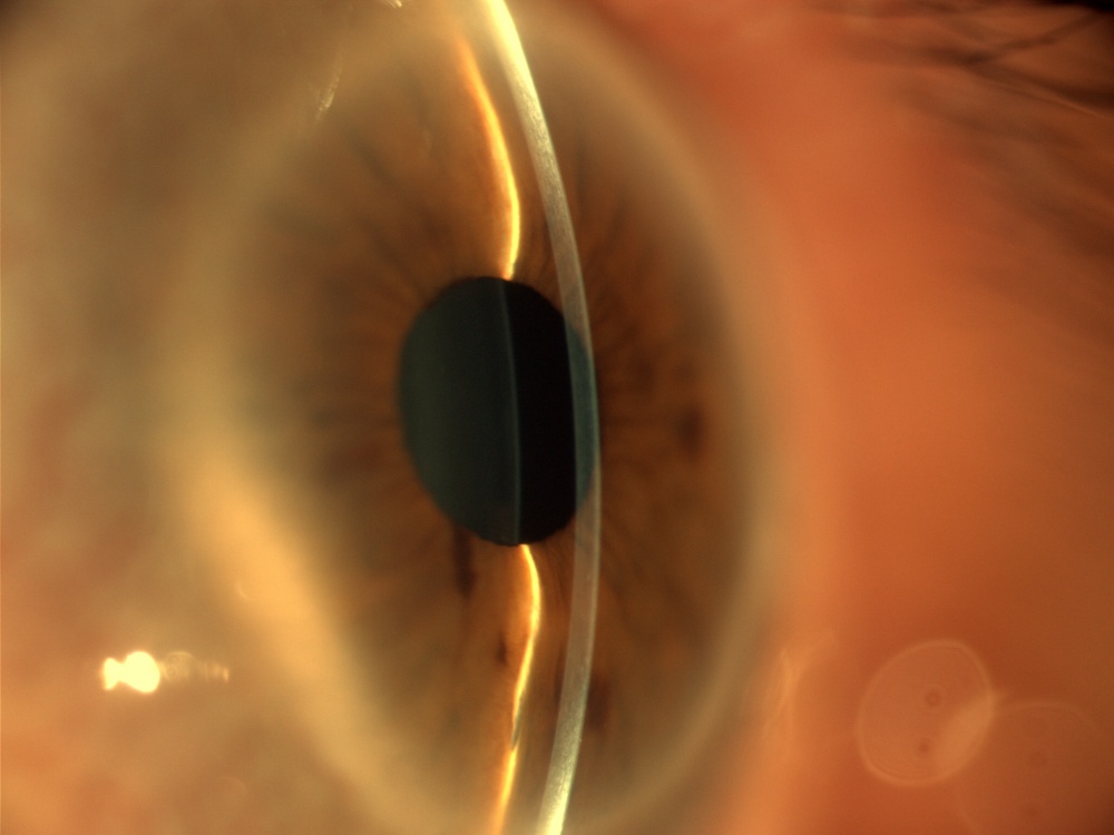
- Right eye corneal thickness measurement of 300 microns (normal range is approximately 550 microns)
- Reduced corneal curvature
- No other significant ocular findings
Diagnosis:
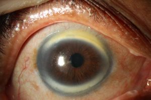
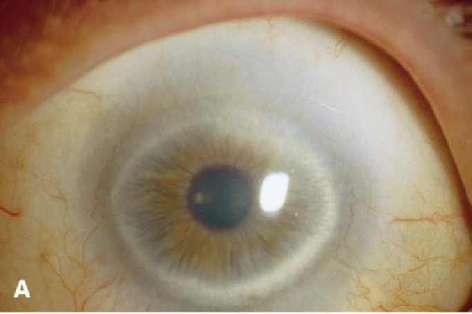
- The diagnosis of cornea plana was confirmed.
cornea plana DISEASE entity
Cornea plana is a congenital condition where the cornea is flattened and the angle between it and the sclera is decreased. This may lead to hyperopia, a hazy corneal limbus, and early development of arcus lipoids.
Cornea plana have both autosomal dominant and autosomal recessive inheritance:
1: Autosomal dominant. CNA1, MIM 121400. Mild disease.
2: Autosomal recessive. CNA2, MIM 217300. Homozygous mutation in the KERA gene. Severe disease. Associated with additional ocular manifestations.
Cornea plana is a rare condition. Families with CNA1 (dominant inheritance) have been studied in Denmark, Germany, the USA, the Netherlands, and Cuba. CNA2 (recessive inheritance) has been identified in Finnish and Saudi peoples.
The locus for CNA2 has been mapped in Finnish families. This locus is in a 3 cM interval with markers D12S82 and D12S327 on either side of chromosome 12.
There is a high prevalence in those in northern Finland and the lower parts of the Kemijoki river in Finland. It affects males and females equally. Finnish cases make up 80% of the CNA2 cases. Saudi Arabia is the second most common background of patients with this disease.
Cornea Plana MANAGEMENT
Cornea plana require management of refractive errors and monitoring for glaucoma over one’s lifetime. Insufficient evidence has been found supporting penetrating keratoplasty.
It is difficult for patients to use contact lenses since they can result in irritation and movement of the eye due to the abnormal shape of the cornea.
When prescribing glasses for a patient, recommend basing these on subjective refraction and giving lenses that are 3-4 D weaker than objective values.
HOW TO TAKE SLIT-LAMP EXAM IMAGES WITH A SMARTPHONE?
Smartphone slit-lamp photography is the new advancement in the field of science and technology in which photographs of the desired slit-lamp finding can be taken with smartphones by using the slit-lamp adapters.
Slit-lamp Smartphone photography
REFERENCES
- Sutton G, Lawless MA, Rogers CM. . Aust N Z J Ophthalmol. 1995;23(1):74-75. doi:10.1111/j.1442-9071.1995.tb01651.x
- National Library of Medicine. Cornea Plana.
- Tahvanainen E, Forsius H, Kolehmainen J, Damsten M, Fellman J, de la Chapelle A. The genetics of cornea plana congenita. J Med Genet. 1996;33(2):116-119. doi:10.1136/jmg.33.2.116
- Forsius H, Damsten M, Eriksson AW, Fellman J, Lindh S, Tahvanainen E. Autosomal recessive. A clinical and genetic study of 78 cases in Finland. Acta Ophthalmol Scand.
- Online Mendelian Inheritance in Man, Autosomal Dominant; CNA1. https://disorders.eyes.arizona.edu/handouts/cornea-plana. Accessed August 10, 2022.
- Sigler-Villanueva A, Tahvanainen E, Lindh S, Dieguez-Lucena J, Forsius H. Autosomal dominant : clinical findings in a Cuban family and a review of the literature. Ophthalmic Genet. 1997;18(2):55-62. doi:10.3109/13816819709057116.
- Online Mendelian Inheritance in Man. Cornea Plana 2, Autosomal Recessive; CNA2.
